 Movie
Movie Controller

[English] 日本語
 Yorodumi
- PDB-2req: METHYLMALONYL-COA MUTASE, NON-PRODUCTIVE COA COMPLEX, IN OPEN CON... -
+ Open data

- Basic information
| Entry | Database: PDB / ID: 2req | ||||||
|---|---|---|---|---|---|---|---|
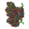
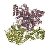
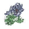
| Title | METHYLMALONYL-COA MUTASE, NON-PRODUCTIVE COA COMPLEX, IN OPEN CONFORMATION REPRESENTING SUBSTRATE-FREE STATE | ||||||
 Components Components | (METHYLMALONYL-COA MUTASE) x 2 | ||||||
 Keywords Keywords | ISOMERASE / MUTASE / INTRAMOLECULAR TRANSFERASE | ||||||
| Function / homology |  Function and homology information Function and homology information: / : / methylmalonyl-CoA mutase / methylmalonyl-CoA mutase activity / cobalamin binding / metal ion binding / cytoplasm Similarity search - Function | ||||||
| Biological species |  Propionibacterium freudenreichii subsp. shermanii (bacteria) Propionibacterium freudenreichii subsp. shermanii (bacteria) | ||||||
| Method |  X-RAY DIFFRACTION / SYNCHROTRON / MOLECULAR REPLACEMENT / Resolution: 2.5 Å X-RAY DIFFRACTION / SYNCHROTRON / MOLECULAR REPLACEMENT / Resolution: 2.5 Å | ||||||
 Authors Authors | Evans, P.R. / Mancia, F. | ||||||
 Citation Citation | Journal: Structure / Year: 1998 Title: Conformational changes on substrate binding to methylmalonyl CoA mutase and new insights into the free radical mechanism. Authors: Mancia, F. / Evans, P.R. #1: Journal: Structure / Year: 1996Title: How Coenzyme B12 Radicals are Generated: The Crystal Structure of Methylmalonyl-Coenzyme a Mutase at 2 A Resolution Authors: Mancia, F. / Keep, N.H. / Nakagawa, A. / Leadlay, P.F. / Mcsweeney, S. / Rasmussen, B. / Bosecke, P. / Diat, O. / Evans, P.R. | ||||||
| History |
|
- Structure visualization
| Structure viewer | Molecule: MolmilJmol/JSmol |
|---|
- Downloads & links
-Download
| PDBx/mmCIF format | 2req.cif.gz | 540.1 KB | Display | PDBx/mmCIF format |
|---|---|---|---|---|
| PDB format | pdb2req.ent.gz | 430.8 KB | Display | PDB format |
| PDBx/mmJSON format | 2req.json.gz | Tree view | PDBx/mmJSON format | |
| Others |  Other downloads Other downloads |
-Validation report
| Arichive directory | https://data.pdbj.org/pub/pdb/validation_reports/re/2reqftp://data.pdbj.org/pub/pdb/validation_reports/re/2req | HTTPS FTP |
|---|
-Related structure data
| Related structure data |  3reqC 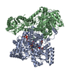 4reqC  1reqS S: Starting model for refinement C: citing same article ( |
|---|---|
| Similar structure data |

-Links
 PDBj
PDBj


- Assembly
| Deposited unit | 
| ||||||||
|---|---|---|---|---|---|---|---|---|---|
| 1 | 
| ||||||||
| 2 | 
| ||||||||
| Unit cell |
| ||||||||
| Noncrystallographic symmetry (NCS) | NCS oper: (Code: given Matrix: (-0.320376, 0.104528, 0.941506), Vector: Details | THE CRYSTAL ASYMMETRIC UNIT CONSISTS OF TWO HETERODIMERIC MOLECULES, EACH WITH AN ACTIVE ALPHA CHAIN (CHAINS A AND C), AND AN INACTIVE BETA CHAIN (CHAINS B AND D). EACH ALPHA CHAIN CONTAINS A COBALAMIN, MODELLED AS A 5-COORDINATE CO(II) SPECIES, AND A COENZYME A MOLECULE APPROXIMATELY IN THE SUBSTRATE BINDING SITE, BUT WITH THE PANTOTHEINE SIDE CHAIN DISORDERED. THE WATERS ARE ASSOCIATED WITH PROTEIN CHAINS AS FOLLOWS: PROTEIN CHAINS WATERS A AND B X C AND D Y | |
-Components
| #1: Protein | Mass: 80137.852 Da / Num. of mol.: 2 Source method: isolated from a genetically manipulated source Details: CHAINS A AND C INCLUDE COENZYME B12, AND COA. B12 IS PRESENT LARGELY AS REDUCED COB(II)ALAMIN, OR B12R. COA IS ONLY PARTLY ORDERED AND IS NOT BOUND CORRECTLY IN THE TRUE SUBSTRATE SITE Source: (gene. exp.) Propionibacterium freudenreichii subsp. shermanii (bacteria)Species: Propionibacterium freudenreichii / Strain: NCIB 9885 Description: THE 2 GENES MUTA (BETA CHAIN) AND MUTB (ALPHA CHAIN) ARE COEXPRESSED FROM THE SAME PLASMID Gene: MUTA, MUTB / Plasmid: PMEX2 / Cellular location (production host): CYTOPLASM / Gene (production host): MUTA, MUTB / Production host: K38 PGP1-2 / References: UniProt: P11653, methylmalonyl-CoA mutase #2: Protein | Mass: 69430.188 Da / Num. of mol.: 2 Source method: isolated from a genetically manipulated source Details: CHAINS A AND C INCLUDE COENZYME B12, AND COA. B12 IS PRESENT LARGELY AS REDUCED COB(II)ALAMIN, OR B12R. COA IS ONLY PARTLY ORDERED AND IS NOT BOUND CORRECTLY IN THE TRUE SUBSTRATE SITE Source: (gene. exp.) Propionibacterium freudenreichii subsp. shermanii (bacteria)Species: Propionibacterium freudenreichii / Strain: NCIB 9885 Description: THE 2 GENES MUTA (BETA CHAIN) AND MUTB (ALPHA CHAIN) ARE COEXPRESSED FROM THE SAME PLASMID Gene: MUTA, MUTB / Plasmid: PMEX2 / Cellular location (production host): CYTOPLASM / Gene (production host): MUTA, MUTB / Production host: K38 PGP1-2 / References: UniProt: P11652, methylmalonyl-CoA mutase #3: Chemical |   Mass: 767.534 Da / Num. of mol.: 2 / Source method: obtained synthetically / Formula: C21H36N7O16P3S Mass: 767.534 Da / Num. of mol.: 2 / Source method: obtained synthetically / Formula: C21H36N7O16P3S#4: Chemical |   Mass: 1330.356 Da / Num. of mol.: 2 / Source method: obtained synthetically / Formula: C62H89CoN13O14P Mass: 1330.356 Da / Num. of mol.: 2 / Source method: obtained synthetically / Formula: C62H89CoN13O14P#5: Water | ChemComp-HOH / |  Mass: 18.015 Da / Num. of mol.: 636 / Source method: isolated from a natural source / Formula: H2O Mass: 18.015 Da / Num. of mol.: 636 / Source method: isolated from a natural source / Formula: H2OCompound details | THIS STRUCTURE IS IN THE OPEN SUBSTRATE-FREE CONFORMATION, WITH THE SUBSTRATE ANALOGUE COA BOUND IN ...THIS STRUCTURE IS IN THE OPEN SUBSTRATE-FREE CONFORMATI | |
|---|
-Experimental details
-Experiment
| Experiment | Method: X-RAY DIFFRACTION / Number of used crystals: 1 |
|---|
- Sample preparation
| Crystal | Density Matthews: 2.8 Å3/Da / Density % sol: 56 % / Description: DATA COLLECTED AT ELETTRA, TRIESTE, ITALY | |||||||||||||||||||||||||||||||||||||||||||||
|---|---|---|---|---|---|---|---|---|---|---|---|---|---|---|---|---|---|---|---|---|---|---|---|---|---|---|---|---|---|---|---|---|---|---|---|---|---|---|---|---|---|---|---|---|---|---|
| Crystal grow | Method: vapor diffusion / pH: 7.5 Details: PROTEIN SOLUTION: 20 MG/ML PROTEIN, 1MM ADENOSYLCOBALAMIN, 2MM COA, 1MM DTT, TRIS -HCL PH 7.5. RESERVOIR SOLUTION: 17.5% (W/V) PEG4000, 20% GLYCEROL (V/V), 100MM TRIS-HCL PH 7.5. EQUAL ...Details: PROTEIN SOLUTION: 20 MG/ML PROTEIN, 1MM ADENOSYLCOBALAMIN, 2MM COA, 1MM DTT, TRIS -HCL PH 7.5. RESERVOIR SOLUTION: 17.5% (W/V) PEG4000, 20% GLYCEROL (V/V), 100MM TRIS-HCL PH 7.5. EQUAL VOLUMES OF PROTEIN SOLUTION WERE MIXED AND EQUILIBRATED BY VAPOR DIFFUSION., vapor diffusion | |||||||||||||||||||||||||||||||||||||||||||||
| Crystal grow | *PLUS Temperature: 23 ℃ / Method: vapor diffusion | |||||||||||||||||||||||||||||||||||||||||||||
| Components of the solutions | *PLUS
|
-Data collection
| Diffraction | Mean temperature: 100 K |
|---|---|
| Diffraction source | Source: SYNCHROTRON / Site: ELETTRA  / Beamline: 5.2R / Wavelength: 1.24 / Beamline: 5.2R / Wavelength: 1.24 |
| Detector | Type: MAR scanner 180 mm plate / Detector: IMAGE PLATE / Date: Jul 1, 1995 / Details: TOROIDAL MIRROR, SI MONOCHROMATOR |
| Radiation | Monochromator: SI(111) / Monochromatic (M) / Laue (L): M / Scattering type: x-ray |
| Radiation wavelength | Wavelength: 1.24 Å / Relative weight: 1 |
| Reflection | Resolution: 2.5→20 Å / Num. obs: 113512 / % possible obs: 99.8 % / Observed criterion σ(I): 3.5 / Redundancy: 4.8 % / Biso Wilson estimate: 55 Å2 / Rmerge(I) obs: 0.092 / Net I/σ(I): 6 |
| Reflection shell | Resolution: 2.5→2.64 Å / Redundancy: 3.7 % / Rmerge(I) obs: 0.338 / Mean I/σ(I) obs: 2.1 / % possible all: 99.7 |
| Reflection shell | *PLUS Highest resolution: 2.5 Å / % possible obs: 99.7 % |
- Processing
| Software |
| ||||||||||||||||||||||||||||||||||||||||||||||||||||||||||||||||||||||||||||||||||||
|---|---|---|---|---|---|---|---|---|---|---|---|---|---|---|---|---|---|---|---|---|---|---|---|---|---|---|---|---|---|---|---|---|---|---|---|---|---|---|---|---|---|---|---|---|---|---|---|---|---|---|---|---|---|---|---|---|---|---|---|---|---|---|---|---|---|---|---|---|---|---|---|---|---|---|---|---|---|---|---|---|---|---|---|---|---|
| Refinement | Method to determine structure: MOLECULAR REPLACEMENT Starting model: PDB ENTRY 1REQ Resolution: 2.5→20 Å / Cross valid method: THROUGHOUT / σ(F): 0
| ||||||||||||||||||||||||||||||||||||||||||||||||||||||||||||||||||||||||||||||||||||
| Displacement parameters | Biso mean: 47 Å2 | ||||||||||||||||||||||||||||||||||||||||||||||||||||||||||||||||||||||||||||||||||||
| Refinement step | Cycle: LAST / Resolution: 2.5→20 Å
| ||||||||||||||||||||||||||||||||||||||||||||||||||||||||||||||||||||||||||||||||||||
| Refine LS restraints |
| ||||||||||||||||||||||||||||||||||||||||||||||||||||||||||||||||||||||||||||||||||||
| Software | *PLUS Name: REFMAC / Classification: refinement | ||||||||||||||||||||||||||||||||||||||||||||||||||||||||||||||||||||||||||||||||||||
| Refinement | *PLUS Num. reflection all: 113303 / Rfactor all: 0.259 | ||||||||||||||||||||||||||||||||||||||||||||||||||||||||||||||||||||||||||||||||||||
| Solvent computation | *PLUS | ||||||||||||||||||||||||||||||||||||||||||||||||||||||||||||||||||||||||||||||||||||
| Displacement parameters | *PLUS |
