 Movie
Movie Controller

[English] 日本語
 Yorodumi
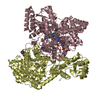
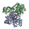
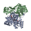
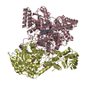
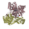
- PDB-6oxd: Structure of Mycobacterium tuberculosis methylmalonyl-CoA mutase ... -
+ Open data

- Basic information
| Entry | Database: PDB / ID: 6oxd | |||||||||
|---|---|---|---|---|---|---|---|---|---|---|
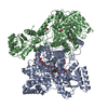
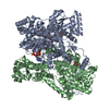
| Title | Structure of Mycobacterium tuberculosis methylmalonyl-CoA mutase with adenosyl cobalamin | |||||||||
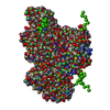
 Components Components | (Methylmalonyl-CoA mutase ...) x 2 | |||||||||
 Keywords Keywords | ISOMERASE / methylmalonyl CoA mutase / methylmalonyl CoA mutase deficiency / metabolic disease / cobalamin / cobalt / disease mutation / metal-binding / itaconyl CoA / radical / Mycobacterium tuberculosis / immunometabolite / B12 / methylmalonic aciduria | |||||||||
| Function / homology |  Function and homology information Function and homology information: / : / methylmalonyl-CoA mutase / methylmalonyl-CoA mutase activity / cobalamin binding / peptidoglycan-based cell wall / metal ion binding / plasma membrane / cytosol / cytoplasm Similarity search - Function | |||||||||
| Biological species |   Mycobacterium tuberculosis (bacteria) Mycobacterium tuberculosis (bacteria) | |||||||||
| Method |  X-RAY DIFFRACTION / SYNCHROTRON / MOLECULAR REPLACEMENT / Resolution: 2 Å X-RAY DIFFRACTION / SYNCHROTRON / MOLECULAR REPLACEMENT / Resolution: 2 Å | |||||||||
 Authors Authors | Purchal, M. / Ruetz, M. / Banerjee, R. / Koutmos, M. | |||||||||
| Funding support |  United States, 2items United States, 2items
| |||||||||
 Citation Citation | Journal: Science / Year: 2019 Title: Itaconyl-CoA forms a stable biradical in methylmalonyl-CoA mutase and derails its activity and repair. Authors: Ruetz, M. / Campanello, G.C. / Purchal, M. / Shen, H. / McDevitt, L. / Gouda, H. / Wakabayashi, S. / Zhu, J. / Rubin, E.J. / Warncke, K. / Mootha, V.K. / Koutmos, M. / Banerjee, R. | |||||||||
| History |
|
- Structure visualization
| Structure viewer | Molecule: MolmilJmol/JSmol |
|---|
- Downloads & links
-Download
| PDBx/mmCIF format | 6oxd.cif.gz | 512.4 KB | Display | PDBx/mmCIF format |
|---|---|---|---|---|
| PDB format | pdb6oxd.ent.gz | 412.5 KB | Display | PDB format |
| PDBx/mmJSON format | 6oxd.json.gz | Tree view | PDBx/mmJSON format | |
| Others |  Other downloads Other downloads |
-Validation report
| Arichive directory | https://data.pdbj.org/pub/pdb/validation_reports/ox/6oxdftp://data.pdbj.org/pub/pdb/validation_reports/ox/6oxd | HTTPS FTP |
|---|
-Related structure data
| Related structure data |  6oxcC 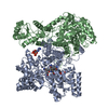 2reqS C: citing same article ( S: Starting model for refinement |
|---|---|
| Similar structure data |

-Links
 PDBj
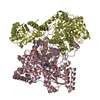
PDBj- Assembly
| Deposited unit | 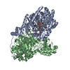
| ||||||||
|---|---|---|---|---|---|---|---|---|---|
| 1 |
| ||||||||
| Unit cell |
|
-Components
-Methylmalonyl-CoA mutase ... , 2 types, 2 molecules AB
| #1: Protein | Mass: 80692.031 Da / Num. of mol.: 1 Source method: isolated from a genetically manipulated source Source: (gene. exp.) Mycobacterium tuberculosis (bacteria)Gene: mutB, DK316_08225, DSI35_00620, ERS027651_01113, ERS027652_00280, ERS124361_00189, SAMEA2682864_01878, SAMEA2683035_01024 Production host: References: UniProt: A0A0E8UW09, UniProt: P9WJK5*PLUS, methylmalonyl-CoA mutase |
|---|---|
| #2: Protein | Mass: 64869.176 Da / Num. of mol.: 1 Source method: isolated from a genetically manipulated source Source: (gene. exp.) Mycobacterium tuberculosis (bacteria)Gene: mutA, DK316_08220, ERS007663_02000, ERS023446_02236, ERS027651_01114, ERS027656_01366, ERS124361_00188, SAMEA2682864_01877, SAMEA2683035_01025 Production host: References: UniProt: A0A045IZR3, UniProt: P9WJK7*PLUS, methylmalonyl-CoA mutase |
-Non-polymers , 5 types, 1124 molecules 


| #3: Chemical | ChemComp-B12 /  Mass: 1330.356 Da / Num. of mol.: 1 / Source method: obtained synthetically / Formula: C62H89CoN13O14P / Feature type: SUBJECT OF INVESTIGATION Mass: 1330.356 Da / Num. of mol.: 1 / Source method: obtained synthetically / Formula: C62H89CoN13O14P / Feature type: SUBJECT OF INVESTIGATION |
|---|---|
| #4: Chemical | ChemComp-5AD /  Mass: 251.242 Da / Num. of mol.: 1 / Source method: obtained synthetically / Formula: C10H13N5O3 / Feature type: SUBJECT OF INVESTIGATION Mass: 251.242 Da / Num. of mol.: 1 / Source method: obtained synthetically / Formula: C10H13N5O3 / Feature type: SUBJECT OF INVESTIGATION |
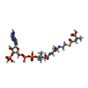
| #5: Chemical | ChemComp-NJS /  Mass: 881.633 Da / Num. of mol.: 1 / Source method: obtained synthetically / Formula: C26H42N7O19P3S / Feature type: SUBJECT OF INVESTIGATION Mass: 881.633 Da / Num. of mol.: 1 / Source method: obtained synthetically / Formula: C26H42N7O19P3S / Feature type: SUBJECT OF INVESTIGATION |
| #6: Chemical | ChemComp-K /  Mass: 39.098 Da / Num. of mol.: 1 / Source method: obtained synthetically / Formula: K Mass: 39.098 Da / Num. of mol.: 1 / Source method: obtained synthetically / Formula: K |
| #7: Water | ChemComp-HOH / Mass: 18.015 Da / Num. of mol.: 1120 / Source method: isolated from a natural source / Formula: H2O |
-Experimental details
-Experiment
| Experiment | Method: X-RAY DIFFRACTION / Number of used crystals: 1 |
|---|
- Sample preparation
| Crystal | Density Matthews: 2.54 Å3/Da / Density % sol: 51.6 % |
|---|---|
| Crystal grow | Temperature: 293 K / Method: vapor diffusion, sitting drop Details: 0.4 M sodium phosphate monobasic, 1.6 M potassium phosphate dibasic, 0.1 M imidazole/HCl, pH 8, 0.2 M sodium chloride |
-Data collection
| Diffraction | Mean temperature: 100 K / Serial crystal experiment: N |
|---|---|
| Diffraction source | Source: SYNCHROTRON / Site: APS / Beamline: 23-ID-D / Wavelength: 1.0332 Å |
| Detector | Type: DECTRIS PILATUS 6M / Detector: PIXEL / Date: Aug 7, 2018 Details: K-B pair of biomorph mirrors for vertical and horizontal focusing |
| Radiation | Monochromator: double crystal Si(111) / Protocol: SINGLE WAVELENGTH / Monochromatic (M) / Laue (L): M / Scattering type: x-ray |
| Radiation wavelength | Wavelength: 1.0332 Å / Relative weight: 1 |
| Reflection | Resolution: 2→29.8 Å / Num. obs: 104126 / % possible obs: 98.4 % / Redundancy: 4.6 % / CC1/2: 0.996 / Rmerge(I) obs: 0.103 / Rrim(I) all: 0.127 / Net I/σ(I): 8.9 |
| Reflection shell | Resolution: 2→2.03 Å / Redundancy: 4.5 % / Rmerge(I) obs: 0.997 / Num. unique obs: 5172 / CC1/2: 0.622 / Rrim(I) all: 1.243 / % possible all: 99.8 |
- Processing
| Software |
| |||||||||||||||||||||||||||||||||||||||||||||||||||||||||||||||||||||||||||||||||||||||||||||||||||||||||||||||||||||||||||||||||||||||||||||||||||||||||||||||||||||||||||||||||||||||||||||||||||||||||||||||||||||||||
|---|---|---|---|---|---|---|---|---|---|---|---|---|---|---|---|---|---|---|---|---|---|---|---|---|---|---|---|---|---|---|---|---|---|---|---|---|---|---|---|---|---|---|---|---|---|---|---|---|---|---|---|---|---|---|---|---|---|---|---|---|---|---|---|---|---|---|---|---|---|---|---|---|---|---|---|---|---|---|---|---|---|---|---|---|---|---|---|---|---|---|---|---|---|---|---|---|---|---|---|---|---|---|---|---|---|---|---|---|---|---|---|---|---|---|---|---|---|---|---|---|---|---|---|---|---|---|---|---|---|---|---|---|---|---|---|---|---|---|---|---|---|---|---|---|---|---|---|---|---|---|---|---|---|---|---|---|---|---|---|---|---|---|---|---|---|---|---|---|---|---|---|---|---|---|---|---|---|---|---|---|---|---|---|---|---|---|---|---|---|---|---|---|---|---|---|---|---|---|---|---|---|---|---|---|---|---|---|---|---|---|---|---|---|---|---|---|---|---|
| Refinement | Method to determine structure: MOLECULAR REPLACEMENT Starting model: PDB entry 2REQ Resolution: 2→29.798 Å / SU ML: 0.22 / Cross valid method: FREE R-VALUE / σ(F): 1.34 / Phase error: 19.9
| |||||||||||||||||||||||||||||||||||||||||||||||||||||||||||||||||||||||||||||||||||||||||||||||||||||||||||||||||||||||||||||||||||||||||||||||||||||||||||||||||||||||||||||||||||||||||||||||||||||||||||||||||||||||||
| Solvent computation | Shrinkage radii: 0.9 Å / VDW probe radii: 1.11 Å | |||||||||||||||||||||||||||||||||||||||||||||||||||||||||||||||||||||||||||||||||||||||||||||||||||||||||||||||||||||||||||||||||||||||||||||||||||||||||||||||||||||||||||||||||||||||||||||||||||||||||||||||||||||||||
| Refinement step | Cycle: LAST / Resolution: 2→29.798 Å
| |||||||||||||||||||||||||||||||||||||||||||||||||||||||||||||||||||||||||||||||||||||||||||||||||||||||||||||||||||||||||||||||||||||||||||||||||||||||||||||||||||||||||||||||||||||||||||||||||||||||||||||||||||||||||
| Refine LS restraints |
| |||||||||||||||||||||||||||||||||||||||||||||||||||||||||||||||||||||||||||||||||||||||||||||||||||||||||||||||||||||||||||||||||||||||||||||||||||||||||||||||||||||||||||||||||||||||||||||||||||||||||||||||||||||||||
| LS refinement shell |
|
