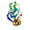
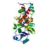
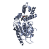
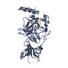
HistoneacetyltransferaseMYST3 / MYST protein 3 / MOZ / YBF2/SAS3 / SAS2 and TIP60 protein 3 / Runt-related transcription factor- ...MYST protein 3 / MOZ / YBF2/SAS3 / SAS2 and TIP60 protein 3 / Runt-related transcription factor-binding protein 2 / Monocytic leukemia zinc finger protein / Zinc finger protein 220
Mass: 34021.637 Da / Num. of mol.: 1 / Fragment: HAT domain Source method: isolated from a genetically manipulated source Source: (gene. exp.) Homo sapiens (human) / Gene: MYST3, MOZ, RUNXBP2, ZNF220 / Production host: Escherichia coli (E. coli) References: UniProt: Q92794, histone acetyltransferase, Transferases; Acyltransferases; Transferring groups other than aminoacyl groups
Av σ(I) over netI: 11.7 / Number: 98811 / Rmerge(I) obs: 0.063 / Χ2: 0.99 / D res high: 3.2 Å / D res low: 50 Å / Num. obs: 13795 / % possible obs: 99.9
Diffraction reflection shell
Highest resolution (Å)
Lowest resolution (Å)
% possible obs (%)
ID
Rmerge(I) obs
Chi squared
6.89
50
99.2
1
0.025
0.876
5.47
6.89
100
1
0.041
1.062
4.78
5.47
100
1
0.045
0.967
4.34
4.78
100
1
0.047
0.955
4.03
4.34
100
1
0.066
0.971
3.79
4.03
100
1
0.109
0.996
3.6
3.79
100
1
0.155
1.057
3.45
3.6
100
1
0.223
0.997
3.31
3.45
100
1
0.327
1.006
3.2
3.31
100
1
0.458
1.075
Reflection
Resolution: 2.67→50 Å / Num. obs: 12178 / % possible obs: 95.2 % / Rmerge(I) obs: 0.082 / Χ2: 1.05 / Net I/σ(I): 13.6
Reflection shell
Resolution (Å)
Rmerge(I) obs
Num. unique all
Χ2
Diffraction-ID
% possible all
2.67-2.77
0.072
778
0.991
1,2
62.3
2.77-2.88
0.654
1185
0.953
1,2
94.3
2.88-3.01
0.479
1248
0.968
1,2
99.9
3.01-3.17
0.269
1250
1.074
1,2
100
3.17-3.36
0.151
1265
1.084
1,2
100
3.36-3.62
0.108
1270
1.048
1,2
100
3.62-3.99
0.08
1275
1.092
1,2
100
3.99-4.56
0.066
1295
1.002
1,2
100
4.56-5.75
0.066
1297
1.091
1,2
99.8
5.75-50
0.067
1315
1.096
1,2
94.7
-
Phasing
Phasing
Method: MAD
Phasing set
ID
1
2
3
Phasing MAD
D res high: 3.5 Å / D res low: 20 Å / FOM : 0.56 / Reflection: 5682
Phasing MAD set
Clust-ID
Expt-ID
Set-ID
Wavelength (Å)
F double prime refined
F prime refined
1
3wavelength
1
1.28
4.76
-2.55
1
3wavelength
2
1.2836
3.41
-8.83
1
3wavelength
3
1.2832
5.91
-10.45
Phasing MAD set site
Atom type symbol: Zn / B iso: 60 / Fract x: 0.119 / Fract y: 0.407 / Fract z: 0.058 / Occupancy: 0.684
In the structure databanks used in Yorodumi, some data are registered as the other names, "COVID-19 virus" and "2019-nCoV". Here are the details of the virus and the list of structure data.
Jan 31, 2019. EMDB accession codes are about to change! (news from PDBe EMDB page)
EMDB accession codes are about to change! (news from PDBe EMDB page)
The allocation of 4 digits for EMDB accession codes will soon come to an end. Whilst these codes will remain in use, new EMDB accession codes will include an additional digit and will expand incrementally as the available range of codes is exhausted. The current 4-digit format prefixed with “EMD-” (i.e. EMD-XXXX) will advance to a 5-digit format (i.e. EMD-XXXXX), and so on. It is currently estimated that the 4-digit codes will be depleted around Spring 2019, at which point the 5-digit format will come into force.
The EM Navigator/Yorodumi systems omit the EMD- prefix.
Related info.:Q: What is EMD? / ID/Accession-code notation in Yorodumi/EM Navigator
Yorodumi is a browser for structure data from EMDB, PDB, SASBDB, etc.
This page is also the successor to EM Navigator detail page, and also detail information page/front-end page for Omokage search.
The word "yorodu" (or yorozu) is an old Japanese word meaning "ten thousand". "mi" (miru) is to see.
Related info.:EMDB / PDB / SASBDB / Comparison of 3 databanks / Yorodumi Search / Aug 31, 2016. New EM Navigator & Yorodumi / Yorodumi Papers / Jmol/JSmol / Function and homology information / Changes in new EM Navigator and Yorodumi
 Movie
Movie Controller


 Open data
 Basic information
 Components
 Keywords
 Function and homology information
 Homo sapiens (human)
 X-RAY DIFFRACTION /
  Authors
 Citation
 Structure visualization
 Downloads & links
 Other downloads
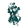


 PDBj
PDBj


 Assembly


 Mass: 65.409 Da / Num. of mol.: 1 / Source method: obtained synthetically / Formula: Zn
 Mass: 809.571 Da / Num. of mol.: 1 / Source method: obtained synthetically / Formula: C23H38N7O17P3S
 Mass: 18.015 Da / Num. of mol.: 12 / Source method: isolated from a natural source / Formula: H2O
 Sample preparation
 / Beamline: 19-BM / Wavelength: 0.9795, 1.2571, 1.2829, 1.2832
 Processing