 Movie
Movie Controller

[English] 日本語
 Yorodumi
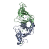
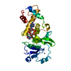
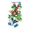
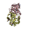
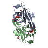
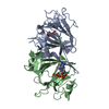
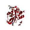
- PDB-3fe5: Crystal structure of 3-hydroxyanthranilate 3,4-dioxygenase from b... -
+ Open data

- Basic information
| Entry | Database: PDB / ID: 3fe5 | ||||||
|---|---|---|---|---|---|---|---|
| Title | Crystal structure of 3-hydroxyanthranilate 3,4-dioxygenase from bovine kidney | ||||||
 Components Components | 3-hydroxyanthranilate 3,4-dioxygenase | ||||||
 Keywords Keywords | OXIDOREDUCTASE / CUPIN / 3HAO / Quinolinic acid / Cytoplasm / Dioxygenase / Iron / Metal-binding | ||||||
| Function / homology |  Function and homology information Function and homology informationquinolinate metabolic process / 3-hydroxyanthranilate 3,4-dioxygenase / 3-hydroxyanthranilate 3,4-dioxygenase activity / : / 'de novo' NAD+ biosynthetic process from L-tryptophan / L-tryptophan catabolic process / quinolinate biosynthetic process / NAD+ biosynthetic process / ferrous iron binding / cytosol / cytoplasm Similarity search - Function | ||||||
| Biological species |  | ||||||
| Method |  X-RAY DIFFRACTION / SYNCHROTRON / MOLECULAR REPLACEMENT / Resolution: 2.51 Å X-RAY DIFFRACTION / SYNCHROTRON / MOLECULAR REPLACEMENT / Resolution: 2.51 Å | ||||||
 Authors Authors | Dilovic, I. / Gliubich, F. / Malpeli, G. / Zanotti, G. / Matkovic-Calogovic, D. | ||||||
 Citation Citation | Journal: Biopolymers / Year: 2009 Title: Crystal structure of bovine 3-hydroxyanthranilate 3,4-dioxygenase. Authors: Dilovic, I. / Gliubich, F. / Malpeli, G. / Zanotti, G. / Matkovic-Calogovic, D. #1: Journal: Biochemistry / Year: 2005Title: Structural studies on 3-hydroxyanthranilate-3,4-dioxygenase: the catalytic mechanism of a complex oxidation involved in NAD biosynthesis. Authors: Zhang, Y. / Colabroy, K.L. / Begley, T.P. / Ealick, S.E. #2: Journal: Protein Sci. / Year: 2006 Title: Crystal structure of 3-hydroxyanthranilic acid 3,4-dioxygenase from Saccharomyces cerevisiae: a special subgroup of the type III extradiol dioxygenases. Authors: Li, X. / Guo, M. / Fan, J. / Tang, W. / Wang, D. / Ge, H. / Rong, H. / Teng, M. / Niu, L. / Liu, Q. / Hao, Q. | ||||||
| History |
|
- Structure visualization
| Structure viewer | Molecule: MolmilJmol/JSmol |
|---|
- Downloads & links
-Download
| PDBx/mmCIF format | 3fe5.cif.gz | 71.8 KB | Display | PDBx/mmCIF format |
|---|---|---|---|---|
| PDB format | pdb3fe5.ent.gz | 52.2 KB | Display | PDB format |
| PDBx/mmJSON format | 3fe5.json.gz | Tree view | PDBx/mmJSON format | |
| Others |  Other downloads Other downloads |
-Validation report
| Arichive directory | https://data.pdbj.org/pub/pdb/validation_reports/fe/3fe5ftp://data.pdbj.org/pub/pdb/validation_reports/fe/3fe5 | HTTPS FTP |
|---|
-Related structure data
| Related structure data |  2qnkS S: Starting model for refinement |
|---|---|
| Similar structure data |


-Links
 PDBj
PDBj- Assembly
| Deposited unit | 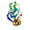
| ||||||||
|---|---|---|---|---|---|---|---|---|---|
| 1 |
| ||||||||
| Unit cell |
|
-Components
| #1: Protein | Mass: 32448.875 Da / Num. of mol.: 1 / Source method: isolated from a natural source / Source: (natural) References: UniProt: Q0VCA8, 3-hydroxyanthranilate 3,4-dioxygenase |
|---|---|
| #2: Chemical | ChemComp-FE /   Mass: 55.845 Da / Num. of mol.: 1 / Source method: obtained synthetically / Formula: Fe Mass: 55.845 Da / Num. of mol.: 1 / Source method: obtained synthetically / Formula: Fe |
| #3: Water | ChemComp-HOH /  Mass: 18.015 Da / Num. of mol.: 55 / Source method: isolated from a natural source / Formula: H2O Mass: 18.015 Da / Num. of mol.: 55 / Source method: isolated from a natural source / Formula: H2O |
| Has protein modification | Y |
-Experimental details
-Experiment
| Experiment | Method: X-RAY DIFFRACTION / Number of used crystals: 2 |
|---|
- Sample preparation
| Crystal | Density Matthews: 2.11 Å3/Da / Density % sol: 41.8 % |
|---|---|
| Crystal grow | Temperature: 298 K / Method: vapor diffusion, sitting drop / pH: 8 Details: 40% (NH4)2SO4, Tris-HCl, 40mM MgCl2, 3% MPD, pH 8, VAPOR DIFFUSION, SITTING DROP, temperature 298K |
-Data collection
| Diffraction | Mean temperature: 100 K |
|---|---|
| Diffraction source | Source: SYNCHROTRON / Site: ELETTRA  / Beamline: 5.2R / Wavelength: 1.2 Å / Beamline: 5.2R / Wavelength: 1.2 Å |
| Detector | Type: MAR scanner 180 mm plate / Detector: IMAGE PLATE / Date: Jul 27, 1996 |
| Radiation | Monochromator: Si (111) / Protocol: SINGLE WAVELENGTH / Monochromatic (M) / Laue (L): M / Scattering type: x-ray |
| Radiation wavelength | Wavelength: 1.2 Å / Relative weight: 1 |
| Reflection | Resolution: 2.51→35.07 Å / Num. all: 8486 / Num. obs: 8486 / % possible obs: 89.7 % / Observed criterion σ(F): 0 / Observed criterion σ(I): 0 / Redundancy: 2.1 % / Rmerge(I) obs: 0.03 / Net I/σ(I): 17.2 |
| Reflection shell | Resolution: 2.51→2.58 Å / Redundancy: 2 % / Rmerge(I) obs: 0.041 / Mean I/σ(I) obs: 3.1 / Num. unique all: 183 / % possible all: 60 |
- Processing
| Software |
| ||||||||||||||||||||||||||||||||||||||||||||||||||||||||||||||||||||||||||||||||||||||||||||||||||||||||||||||||||||||||||||||||||||||||||||||||||||||||||||||||||||||||||
|---|---|---|---|---|---|---|---|---|---|---|---|---|---|---|---|---|---|---|---|---|---|---|---|---|---|---|---|---|---|---|---|---|---|---|---|---|---|---|---|---|---|---|---|---|---|---|---|---|---|---|---|---|---|---|---|---|---|---|---|---|---|---|---|---|---|---|---|---|---|---|---|---|---|---|---|---|---|---|---|---|---|---|---|---|---|---|---|---|---|---|---|---|---|---|---|---|---|---|---|---|---|---|---|---|---|---|---|---|---|---|---|---|---|---|---|---|---|---|---|---|---|---|---|---|---|---|---|---|---|---|---|---|---|---|---|---|---|---|---|---|---|---|---|---|---|---|---|---|---|---|---|---|---|---|---|---|---|---|---|---|---|---|---|---|---|---|---|---|---|---|---|
| Refinement | Method to determine structure: MOLECULAR REPLACEMENT Starting model: 2QNK Resolution: 2.51→35.07 Å / Cor.coef. Fo:Fc: 0.947 / Cor.coef. Fo:Fc free: 0.87 / SU B: 11.378 / SU ML: 0.252 / Cross valid method: THROUGHOUT / σ(F): 0 / σ(I): 0 / ESU R Free: 0.351 / Stereochemistry target values: MAXIMUM LIKELIHOOD / Details: HYDROGENS HAVE BEEN ADDED IN THE RIDING POSITIONS
| ||||||||||||||||||||||||||||||||||||||||||||||||||||||||||||||||||||||||||||||||||||||||||||||||||||||||||||||||||||||||||||||||||||||||||||||||||||||||||||||||||||||||||
| Solvent computation | Ion probe radii: 0.8 Å / Shrinkage radii: 0.8 Å / VDW probe radii: 1.2 Å / Solvent model: MASK | ||||||||||||||||||||||||||||||||||||||||||||||||||||||||||||||||||||||||||||||||||||||||||||||||||||||||||||||||||||||||||||||||||||||||||||||||||||||||||||||||||||||||||
| Displacement parameters | Biso mean: 20.254 Å2
| ||||||||||||||||||||||||||||||||||||||||||||||||||||||||||||||||||||||||||||||||||||||||||||||||||||||||||||||||||||||||||||||||||||||||||||||||||||||||||||||||||||||||||
| Refinement step | Cycle: LAST / Resolution: 2.51→35.07 Å
| ||||||||||||||||||||||||||||||||||||||||||||||||||||||||||||||||||||||||||||||||||||||||||||||||||||||||||||||||||||||||||||||||||||||||||||||||||||||||||||||||||||||||||
| Refine LS restraints |
| ||||||||||||||||||||||||||||||||||||||||||||||||||||||||||||||||||||||||||||||||||||||||||||||||||||||||||||||||||||||||||||||||||||||||||||||||||||||||||||||||||||||||||
| LS refinement shell | Resolution: 2.515→2.58 Å / Total num. of bins used: 20
|
