 Movie
Movie Controller

[English] 日本語
 Yorodumi
- PDB-2rbg: Crystal structure of hypothetical protein(ST0493) from sulfolobus... -
+ Open data

- Basic information
| Entry | Database: PDB / ID: 2rbg | ||||||
|---|---|---|---|---|---|---|---|
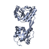
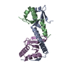
| Title | Crystal structure of hypothetical protein(ST0493) from sulfolobus tokodaii | ||||||
 Components Components | Putative uncharacterized protein ST0493 | ||||||
 Keywords Keywords | STRUCTURAL GENOMICS / UNKNOWN FUNCTION / Hypothetical protein / NPPSFA / National Project on Protein Structural and Functional Analyses / RIKEN Structural Genomics/Proteomics Initiative / RSGI | ||||||
| Function / homology | Rossmann fold - #11100 / Domain of unknown function DUF5751 / Family of unknown function (DUF5751) / Rossmann fold / 3-Layer(aba) Sandwich / Alpha Beta / DUF5751 domain-containing protein Function and homology information Function and homology information | ||||||
| Biological species |   Sulfolobus tokodaii (archaea) Sulfolobus tokodaii (archaea) | ||||||
| Method |  X-RAY DIFFRACTION / SYNCHROTRON / MAD / Resolution: 1.75 Å X-RAY DIFFRACTION / SYNCHROTRON / MAD / Resolution: 1.75 Å | ||||||
 Authors Authors | Jeyakanthan, J. / Kuramitsu, S. / Yokoyama, S. / RIKEN Structural Genomics/Proteomics Initiative (RSGI) | ||||||
 Citation Citation | Journal: To be Published Title: Crystal structure of hypothetical protein(ST0493) from sulfolobus tokodaii Authors: Jeyakanthan, J. / Kuramitsu, S. / Yokoyama, S. | ||||||
| History |
|
- Structure visualization
| Structure viewer | Molecule: MolmilJmol/JSmol |
|---|
- Downloads & links
-Download
| PDBx/mmCIF format | 2rbg.cif.gz | 70.2 KB | Display | PDBx/mmCIF format |
|---|---|---|---|---|
| PDB format | pdb2rbg.ent.gz | 52.1 KB | Display | PDB format |
| PDBx/mmJSON format | 2rbg.json.gz | Tree view | PDBx/mmJSON format | |
| Others |  Other downloads Other downloads |
-Validation report
| Summary document | 2rbg_validation.pdf.gz | 439.4 KB | Display | wwPDB validaton report |
|---|---|---|---|---|
| Full document | 2rbg_full_validation.pdf.gz | 440.1 KB | Display | |
| Data in XML | 2rbg_validation.xml.gz | 16.8 KB | Display | |
| Data in CIF | 2rbg_validation.cif.gz | 23.4 KB | Display | |
| Arichive directory | https://data.pdbj.org/pub/pdb/validation_reports/rb/2rbgftp://data.pdbj.org/pub/pdb/validation_reports/rb/2rbg | HTTPS FTP |
-Related structure data
| Similar structure data | |
|---|---|
| Other databases |


-Links
 PDBj
PDBj- Assembly
| Deposited unit | 
| ||||||||
|---|---|---|---|---|---|---|---|---|---|
| 1 |
| ||||||||
| 2 | 
| ||||||||
| 3 | 
| ||||||||
| Unit cell |
|
-Components
| #1: Protein | Mass: 14961.982 Da / Num. of mol.: 2 Source method: isolated from a genetically manipulated source Source: (gene. exp.) Sulfolobus tokodaii (archaea) / Strain: Strain 7 / Plasmid: pET-21a / Production host:  #2: Chemical | ChemComp-SO4 / |   Mass: 96.063 Da / Num. of mol.: 1 / Source method: obtained synthetically / Formula: SO4 Mass: 96.063 Da / Num. of mol.: 1 / Source method: obtained synthetically / Formula: SO4#3: Water | ChemComp-HOH / |  Mass: 18.015 Da / Num. of mol.: 316 / Source method: isolated from a natural source / Formula: H2O Mass: 18.015 Da / Num. of mol.: 316 / Source method: isolated from a natural source / Formula: H2OHas protein modification | Y | |
|---|
-Experimental details
-Experiment
| Experiment | Method: X-RAY DIFFRACTION / Number of used crystals: 1 |
|---|
- Sample preparation
| Crystal | Density Matthews: 2.11 Å3/Da / Density % sol: 41.69 % |
|---|---|
| Crystal grow | Temperature: 293 K / Method: microbatch / pH: 7.5 Details: 30% PEG 4K, 0.2M Ammonium Sulfate, pH 7.5, MICROBATCH, temperature 293K |
-Data collection
| Diffraction | Mean temperature: 100 K | ||||||||||||
|---|---|---|---|---|---|---|---|---|---|---|---|---|---|
| Diffraction source | Source: SYNCHROTRON / Site: SPring-8  / Beamline: BL26B2 / Wavelength: 0.97899, 0.9, 0.97931 / Beamline: BL26B2 / Wavelength: 0.97899, 0.9, 0.97931 | ||||||||||||
| Detector | Type: MARMOSAIC 225 mm CCD / Detector: CCD / Date: Jun 16, 2007 / Details: RH COATED BENT-CYRINDRICAL MIRROR | ||||||||||||
| Radiation | Monochromator: SI-1 1 1 DOUBLE CRYSTAL MONOCHROMATOR / Protocol: MAD / Monochromatic (M) / Laue (L): M / Scattering type: x-ray | ||||||||||||
| Radiation wavelength |
| ||||||||||||
| Reflection | Resolution: 1.75→50 Å / Num. obs: 25105 / % possible obs: 99.6 % / Biso Wilson estimate: 13.3 Å2 / Rmerge(I) obs: 0.059 / Rsym value: 0.063 | ||||||||||||
| Reflection shell | Resolution: 1.75→1.81 Å / Rmerge(I) obs: 0.143 / Num. unique all: 2433 / Rsym value: 0.133 / % possible all: 96.9 |
- Processing
| Software |
| ||||||||||||||||||||
|---|---|---|---|---|---|---|---|---|---|---|---|---|---|---|---|---|---|---|---|---|---|
| Refinement | Method to determine structure: MAD / Resolution: 1.75→33.49 Å / Rfactor Rfree error: 0.006 / Data cutoff high absF: 2067291.84 / Data cutoff low absF: 0 / Isotropic thermal model: RESTRAINED / Cross valid method: THROUGHOUT / σ(F): 0 / Stereochemistry target values: Engh & Huber
| ||||||||||||||||||||
| Solvent computation | Solvent model: FLAT MODEL / Bsol: 51.2041 Å2 / ksol: 0.371373 e/Å3 | ||||||||||||||||||||
| Displacement parameters | Biso mean: 16.9 Å2
| ||||||||||||||||||||
| Refine analyze |
| ||||||||||||||||||||
| Refinement step | Cycle: LAST / Resolution: 1.75→33.49 Å
| ||||||||||||||||||||
| Refine LS restraints |
| ||||||||||||||||||||
| LS refinement shell | Resolution: 1.75→1.83 Å / Rfactor Rfree error: 0.019 / Total num. of bins used: 8
| ||||||||||||||||||||
| Xplor file |
|
