Type: MARMOSAIC 225 mm CCD / Detector: CCD / Date: Aug 25, 2014 / Details: beryllium lenses
Radiation
Monochromator: C(111) / Protocol: SINGLE WAVELENGTH / Monochromatic (M) / Laue (L): M / Scattering type: x-ray
Radiation wavelength
Wavelength: 0.97872 Å / Relative weight: 1
Reflection
Resolution: 2→50 Å / Num. obs: 9939 / % possible obs: 100 % / Redundancy: 8.6 % / Rmerge(I) obs: 0.095 / Net I/σ(I): 20.1
Reflection shell
Resolution: 2→2.03 Å / Redundancy: 5.8 % / Rmerge(I) obs: 0.61 / Mean I/σ(I) obs: 2.7 / % possible all: 100
-
Processing
Software
Name
Version
Classification
REFMAC
5.8.0135
refinement
HKL-2000
datascaling
PHENIX
modelbuilding
BALBES
phasing
HKL-2000
datareduction
Refinement
Resolution: 2→50 Å / Cor.coef. Fo:Fc: 0.965 / Cor.coef. Fo:Fc free: 0.925 / SU B: 8.573 / SU ML: 0.118 / Cross valid method: THROUGHOUT / ESU R: 0.164 / ESU R Free: 0.165 / Details: HYDROGENS HAVE BEEN ADDED IN THE RIDING POSITIONS
Rfactor
Num. reflection
% reflection
Selection details
Rfree
0.23775
497
5 %
RANDOM
Rwork
0.17262
-
-
-
obs
0.17589
9421
99.83 %
-
Solvent computation
Ion probe radii: 0.8 Å / Shrinkage radii: 0.8 Å / VDW probe radii: 1.2 Å
 Movie
Movie Controller

 Yorodumi
 Open data
 Basic information
 Components
 Keywords
 Function and homology information
 X-RAY DIFFRACTION /
  Authors
 Citation
 Structure visualization
 Downloads & links
 Other downloads


 PDBj
PDBj

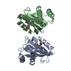
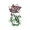
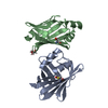
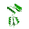
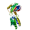
 Assembly
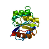

 Mass: 18.015 Da / Num. of mol.: 81 / Source method: isolated from a natural source / Formula: H2O
 Sample preparation
 / Beamline: 21-ID-F / Wavelength: 0.97872 Å
 Processing