Resolution: 2.12→45.175 Å / Num. obs: 14544 / % possible obs: 99.1 % / Observed criterion σ(I): -3 / Biso Wilson estimate: 56.283 Å2 / Rmerge(I) obs: 0.066 / Net I/σ(I): 16.76
Reflection shell
Resolution (Å)
Rmerge(I) obs
Mean I/σ(I) obs
Num. measured obs
Num. unique obs
Diffraction-ID
% possible all
2.12-2.2
0.61
2.3
5155
1480
1,2
99.7
2.2-2.28
0.485
3
4395
1277
1,2
99.3
2.28-2.39
0.356
4
5258
1527
1,2
99.8
2.39-2.51
0.275
5.1
4689
1364
1,2
100
2.51-2.67
0.203
7.1
4967
1448
1,2
99.4
2.67-2.88
0.395
11
7883
1484
1,2
99.6
2.88-3.16
0.285
16.9
9690
1393
1,2
99.1
3.16-3.62
0.104
27.9
10144
1482
1,2
99.1
3.62-4.55
0.044
39.5
9909
1487
1,2
98.8
4.55-45.175
0.029
45.8
9769
1602
1,2
96.7
-
Phasing
Phasing
Method: MAD
-
Processing
Software
Name
Version
Classification
NB
REFMAC
5.2.0019
refinement
PHENIX
refinement
SHELX
phasing
MolProbity
3beta29
modelbuilding
XSCALE
datascaling
PDB_EXTRACT
3
dataextraction
ADSC
Quantum
datacollection
XDS
datareduction
SHELXD
phasing
autoSHARP
phasing
Refinement
Method to determine structure: MAD / Resolution: 2.12→45.175 Å / Cor.coef. Fo:Fc: 0.958 / Cor.coef. Fo:Fc free: 0.942 / SU B: 15.077 / SU ML: 0.172 / TLS residual ADP flag: LIKELY RESIDUAL / Cross valid method: THROUGHOUT / σ(F): 0 / ESU R: 0.193 / ESU R Free: 0.173 Stereochemistry target values: MAXIMUM LIKELIHOOD WITH PHASES Details: 1. HYDROGENS HAVE BEEN ADDED IN THE RIDING POSITIONS. 2. ATOM RECORDS CONTAIN RESIDUAL B FACTORS ONLY. 3. A MET-INHIBITION PROTOCOL WAS USED FOR SELENOMETHIONINE INCORPORATION DURING PROTEIN ...Details: 1. HYDROGENS HAVE BEEN ADDED IN THE RIDING POSITIONS. 2. ATOM RECORDS CONTAIN RESIDUAL B FACTORS ONLY. 3. A MET-INHIBITION PROTOCOL WAS USED FOR SELENOMETHIONINE INCORPORATION DURING PROTEIN EXPRESSION. THE OCCUPANCY OF THE SE ATOMS IN THE MSE RESIDUES WAS REDUCED TO 0.75 FOR THE REDUCED SCATTERING POWER DUE TO PARTIAL S-MET INCORPORATION. 4. SO4 IONS FROM THE CRYSTALLIZATION SOLUTION ARE MODELED. 5. THE FOLLOWING LOOPS HAVE POOR OR NO DENSITY: 5-7, 43-50, 62-69, 165-169. THUS, THE MODEL IN THESE REGIONS IS NOT RELIABLE. N-TERMINUS AS WELL AS PURIFICATION TAG ARE DISORDERED. 6. A FEW NON-WATER DENSITY BLOBS OUTSIDE THE PROTEIN ARE LEFT UNINTERPRETED.
Rfactor
Num. reflection
% reflection
Selection details
Rfree
0.251
699
4.8 %
RANDOM
Rwork
0.215
-
-
-
obs
0.217
14517
99.37 %
-
Solvent computation
Ion probe radii: 0.8 Å / Shrinkage radii: 0.8 Å / VDW probe radii: 1.2 Å / Solvent model: BABINET MODEL WITH MASK
Displacement parameters
Biso mean: 49.003 Å2
Baniso -1
Baniso -2
Baniso -3
1-
1.79 Å2
0 Å2
0 Å2
2-
-
1.79 Å2
0 Å2
3-
-
-
-3.58 Å2
Refinement step
Cycle: LAST / Resolution: 2.12→45.175 Å
Protein
Nucleic acid
Ligand
Solvent
Total
Num. atoms
1349
0
10
27
1386
Refine LS restraints
Refine-ID
Type
Dev ideal
Dev ideal target
Number
X-RAY DIFFRACTION
r_bond_refined_d
0.017
0.022
1402
X-RAY DIFFRACTION
r_bond_other_d
0.002
0.02
955
X-RAY DIFFRACTION
r_angle_refined_deg
1.69
1.976
1898
X-RAY DIFFRACTION
r_angle_other_deg
0.971
3
2322
X-RAY DIFFRACTION
r_dihedral_angle_1_deg
5.093
5
180
X-RAY DIFFRACTION
r_dihedral_angle_2_deg
35.026
23.81
63
X-RAY DIFFRACTION
r_dihedral_angle_3_deg
16.36
15
236
X-RAY DIFFRACTION
r_dihedral_angle_4_deg
16.475
15
12
X-RAY DIFFRACTION
r_chiral_restr
0.105
0.2
214
X-RAY DIFFRACTION
r_gen_planes_refined
0.005
0.02
1563
X-RAY DIFFRACTION
r_gen_planes_other
0.001
0.02
284
X-RAY DIFFRACTION
r_nbd_refined
0.229
0.2
298
X-RAY DIFFRACTION
r_nbd_other
0.203
0.2
972
X-RAY DIFFRACTION
r_nbtor_refined
0.186
0.2
675
X-RAY DIFFRACTION
r_nbtor_other
0.095
0.2
773
X-RAY DIFFRACTION
r_xyhbond_nbd_refined
0.128
0.2
36
X-RAY DIFFRACTION
r_symmetry_vdw_refined
0.135
0.2
8
X-RAY DIFFRACTION
r_symmetry_vdw_other
0.22
0.2
21
X-RAY DIFFRACTION
r_symmetry_hbond_refined
0.157
0.2
4
X-RAY DIFFRACTION
r_mcbond_it
2.409
3
929
X-RAY DIFFRACTION
r_mcbond_other
0.515
3
365
X-RAY DIFFRACTION
r_mcangle_it
3.537
5
1422
X-RAY DIFFRACTION
r_scbond_it
5.965
8
547
X-RAY DIFFRACTION
r_scangle_it
8.748
11
473
LS refinement shell
Resolution: 2.12→2.175 Å / Total num. of bins used: 20
Rfactor
Num. reflection
% reflection
Rfree
0.373
57
-
Rwork
0.312
964
-
all
-
1021
-
obs
-
-
99.71 %
Refinement TLS params.
Method: refined / Origin x: 12.2125 Å / Origin y: 18.0783 Å / Origin z: 57.1044 Å
11
12
13
21
22
23
31
32
33
T
-0.2013 Å2
0.0181 Å2
0.025 Å2
-
0.0018 Å2
-0.0178 Å2
-
-
-0.0102 Å2
L
1.9677 °2
1.1244 °2
1.965 °2
-
2.2797 °2
1.9853 °2
-
-
4.4504 °2
S
0.0767 Å °
0.3603 Å °
-0.1098 Å °
0.0063 Å °
0.176 Å °
0.0019 Å °
-0.1645 Å °
0.1088 Å °
-0.2527 Å °
+
About Yorodumi
-
News
-
Feb 9, 2022. New format data for meta-information of EMDB entries
New format data for meta-information of EMDB entries
Version 3 of the EMDB header file is now the official format.
The previous official version 1.9 will be removed from the archive.
In the structure databanks used in Yorodumi, some data are registered as the other names, "COVID-19 virus" and "2019-nCoV". Here are the details of the virus and the list of structure data.
Jan 31, 2019. EMDB accession codes are about to change! (news from PDBe EMDB page)
EMDB accession codes are about to change! (news from PDBe EMDB page)
The allocation of 4 digits for EMDB accession codes will soon come to an end. Whilst these codes will remain in use, new EMDB accession codes will include an additional digit and will expand incrementally as the available range of codes is exhausted. The current 4-digit format prefixed with “EMD-” (i.e. EMD-XXXX) will advance to a 5-digit format (i.e. EMD-XXXXX), and so on. It is currently estimated that the 4-digit codes will be depleted around Spring 2019, at which point the 5-digit format will come into force.
The EM Navigator/Yorodumi systems omit the EMD- prefix.
Related info.:Q: What is EMD? / ID/Accession-code notation in Yorodumi/EM Navigator
Yorodumi is a browser for structure data from EMDB, PDB, SASBDB, etc.
This page is also the successor to EM Navigator detail page, and also detail information page/front-end page for Omokage search.
The word "yorodu" (or yorozu) is an old Japanese word meaning "ten thousand". "mi" (miru) is to see.
Related info.:EMDB / PDB / SASBDB / Comparison of 3 databanks / Yorodumi Search / Aug 31, 2016. New EM Navigator & Yorodumi / Yorodumi Papers / Jmol/JSmol / Function and homology information / Changes in new EM Navigator and Yorodumi
 Movie
Movie Controller

 Yorodumi
 Open data
 Basic information
 Components
 Keywords
 Function and homology information
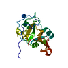
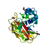
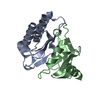
 Thermotoga maritima MSB8 (bacteria)
 X-RAY DIFFRACTION /
  Authors
 Citation
 Structure visualization
 Downloads & links
 Other downloads


 PDBj
PDBj Assembly

 Mass: 96.063 Da / Num. of mol.: 2 / Source method: obtained synthetically / Formula: SO4
 Mass: 18.015 Da / Num. of mol.: 27 / Source method: isolated from a natural source / Formula: H2O
 Sample preparation
 Processing