 Movie
Movie Controller

[English] 日本語
 Yorodumi
- PDB-3q6z: HUman PARP14 (ARTD8)-Macro domain 1 in complex with adenosine-5-d... -
+ Open data

- Basic information
| Entry | Database: PDB / ID: 3q6z | ||||||
|---|---|---|---|---|---|---|---|
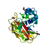
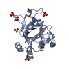
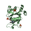
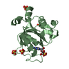
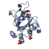
| Title | HUman PARP14 (ARTD8)-Macro domain 1 in complex with adenosine-5-diphosphoribose | ||||||
 Components Components | Poly [ADP-ribose] polymerase 14 | ||||||
 Keywords Keywords | TRANSFERASE / Structural Genomics Consortium / SGC / Adp-ribose binding | ||||||
| Function / homology |  Function and homology information Function and homology informationnegative regulation of tyrosine phosphorylation of STAT protein / positive regulation of interleukin-4-mediated signaling pathway / NAD+ catabolic process / Maturation of nucleoprotein / Nicotinate metabolism / positive regulation of tyrosine phosphorylation of STAT protein / Maturation of nucleoprotein / protein poly-ADP-ribosylation / NAD+-protein-glutamate ADP-ribosyltransferase activity / negative regulation of type II interferon-mediated signaling pathway ...negative regulation of tyrosine phosphorylation of STAT protein / positive regulation of interleukin-4-mediated signaling pathway / NAD+ catabolic process / Maturation of nucleoprotein / Nicotinate metabolism / positive regulation of tyrosine phosphorylation of STAT protein / Maturation of nucleoprotein / protein poly-ADP-ribosylation / NAD+-protein-glutamate ADP-ribosyltransferase activity / negative regulation of type II interferon-mediated signaling pathway / NAD+-protein mono-ADP-ribosyltransferase activity / Transferases; Glycosyltransferases; Pentosyltransferases / NAD+ poly-ADP-ribosyltransferase activity / NAD+ binding / nucleotidyltransferase activity / transcription corepressor activity / negative regulation of gene expression / innate immune response / enzyme binding / membrane / nucleus / plasma membrane / cytoplasm / cytosol Similarity search - Function | ||||||
| Biological species |  Homo sapiens (human) Homo sapiens (human) | ||||||
| Method |  X-RAY DIFFRACTION / SYNCHROTRON / MOLECULAR REPLACEMENT / Resolution: 2.23 Å X-RAY DIFFRACTION / SYNCHROTRON / MOLECULAR REPLACEMENT / Resolution: 2.23 Å | ||||||
 Authors Authors | Karlberg, T. / Siponen, M.I. / Arrowsmith, C.H. / Berglund, H. / Bountra, C. / Collins, R. / Edwards, A.M. / Ekblad, T. / Flodin, S. / Flores, A. ...Karlberg, T. / Siponen, M.I. / Arrowsmith, C.H. / Berglund, H. / Bountra, C. / Collins, R. / Edwards, A.M. / Ekblad, T. / Flodin, S. / Flores, A. / Graslund, S. / Kotenyova, T. / Kouznetsova, E. / Moche, M. / Nordlund, P. / Nyman, T. / Persson, C. / Sehic, A. / Thorsell, A.G. / Tresaugues, L. / Wahlberg, E. / Weigelt, J. / Welin, M. / Schuler, H. / Structural Genomics Consortium (SGC) | ||||||
 Citation Citation | Journal: Structure / Year: 2013 Title: Recognition of Mono-ADP-Ribosylated ARTD10 Substrates by ARTD8 Macrodomains. Authors: Forst, A.H. / Karlberg, T. / Herzog, N. / Thorsell, A.G. / Gross, A. / Feijs, K.L. / Verheugd, P. / Kursula, P. / Nijmeijer, B. / Kremmer, E. / Kleine, H. / Ladurner, A.G. / Schuler, H. / Luscher, B. | ||||||
| History |
|
- Structure visualization
| Structure viewer | Molecule: MolmilJmol/JSmol |
|---|
- Downloads & links
-Download
| PDBx/mmCIF format | 3q6z.cif.gz | 53 KB | Display | PDBx/mmCIF format |
|---|---|---|---|---|
| PDB format | pdb3q6z.ent.gz | 36.4 KB | Display | PDB format |
| PDBx/mmJSON format | 3q6z.json.gz | Tree view | PDBx/mmJSON format | |
| Others |  Other downloads Other downloads |
-Validation report
| Arichive directory | https://data.pdbj.org/pub/pdb/validation_reports/q6/3q6zftp://data.pdbj.org/pub/pdb/validation_reports/q6/3q6z | HTTPS FTP |
|---|
-Related structure data
| Related structure data |  3q71C  3v2bC 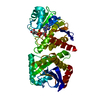 3vfqC 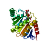 4abkC  4ablC  4d86C 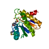 1spvS C: citing same article ( S: Starting model for refinement |
|---|---|
| Similar structure data |


-Links
 PDBj
PDBj
- Assembly
| Deposited unit | 
| ||||||||
|---|---|---|---|---|---|---|---|---|---|
| 1 |
| ||||||||
| Unit cell |
|
-Components
| #1: Protein | Mass: 23514.178 Da / Num. of mol.: 1 / Fragment: MACRO DOMAIN 1, residues 708-898 Source method: isolated from a genetically manipulated source Source: (gene. exp.) Homo sapiens (human) / Gene: BAL2, KIAA1268, PARP14 / Plasmid: pNIC-Bsa4 / Production host:  |
|---|---|
| #2: Chemical | ChemComp-APR /   Mass: 559.316 Da / Num. of mol.: 1 / Source method: obtained synthetically / Formula: C15H23N5O14P2 Mass: 559.316 Da / Num. of mol.: 1 / Source method: obtained synthetically / Formula: C15H23N5O14P2 |
| #3: Water | ChemComp-HOH /  Mass: 18.015 Da / Num. of mol.: 60 / Source method: isolated from a natural source / Formula: H2O Mass: 18.015 Da / Num. of mol.: 60 / Source method: isolated from a natural source / Formula: H2O |
-Experimental details
-Experiment
| Experiment | Method: X-RAY DIFFRACTION / Number of used crystals: 1 |
|---|
- Sample preparation
| Crystal | Density Matthews: 1.93 Å3/Da / Density % sol: 36.39 % |
|---|---|
| Crystal grow | Temperature: 293 K / pH: 8.5 Details: 30% PEGMME2000, 0.1M HEPES, 4mM ADP-RIBOSE, 4mM MAGNESIUM CHLORIDE, pH 8.5, VAPOR DIFFUSION, SITTING DROP, temperature 293K |
-Data collection
| Diffraction | Mean temperature: 100 K |
|---|---|
| Diffraction source | Source: SYNCHROTRON / Site: BESSY  / Beamline: 14.1 / Wavelength: 0.91841 / Beamline: 14.1 / Wavelength: 0.91841 |
| Detector | Type: RAYONIX MX-225 / Detector: CCD / Date: Jan 28, 2010 / Details: MIRRORS |
| Radiation | Monochromator: DOUBLE CRYSTAL MONOCHROMATOR, SI -111 / Protocol: SINGLE WAVELENGTH / Monochromatic (M) / Laue (L): M / Scattering type: x-ray |
| Radiation wavelength | Wavelength: 0.91841 Å / Relative weight: 1 |
| Reflection | Resolution: 2.23→35 Å / Num. obs: 9427 / % possible obs: 99.8 % / Observed criterion σ(I): 0 / Redundancy: 11.8 % / Biso Wilson estimate: 30.5 Å2 / Rmerge(I) obs: 0.093 / Rsym value: 0.079 / Net I/σ(I): 24.8 |
| Reflection shell | Resolution: 2.23→2.29 Å / Redundancy: 12 % / Rmerge(I) obs: 0.623 / Mean I/σ(I) obs: 5.5 / Num. unique all: 667 / Rsym value: 0.325 / % possible all: 99.6 |
- Processing
| Software |
| ||||||||||||||||||||||||||||||||||||||||||||||||||||||||||||||||||||||||||||||||||||||||||||||||||||||||||||||||||||||||||||||||||||||||||||||||||||||||||||||||||||||||||
|---|---|---|---|---|---|---|---|---|---|---|---|---|---|---|---|---|---|---|---|---|---|---|---|---|---|---|---|---|---|---|---|---|---|---|---|---|---|---|---|---|---|---|---|---|---|---|---|---|---|---|---|---|---|---|---|---|---|---|---|---|---|---|---|---|---|---|---|---|---|---|---|---|---|---|---|---|---|---|---|---|---|---|---|---|---|---|---|---|---|---|---|---|---|---|---|---|---|---|---|---|---|---|---|---|---|---|---|---|---|---|---|---|---|---|---|---|---|---|---|---|---|---|---|---|---|---|---|---|---|---|---|---|---|---|---|---|---|---|---|---|---|---|---|---|---|---|---|---|---|---|---|---|---|---|---|---|---|---|---|---|---|---|---|---|---|---|---|---|---|---|---|
| Refinement | Method to determine structure: MOLECULAR REPLACEMENT Starting model: PDB ENTRY 1SPV Resolution: 2.23→31.92 Å / Cor.coef. Fo:Fc: 0.936 / Cor.coef. Fo:Fc free: 0.888 / SU B: 7.2 / SU ML: 0.183 / Cross valid method: THROUGHOUT / σ(F): 0 / ESU R Free: 0.26 / Stereochemistry target values: MAXIMUM LIKELIHOOD / Details: HYDROGENS HAVE BEEN ADDED IN THE RIDING POSITIONS
| ||||||||||||||||||||||||||||||||||||||||||||||||||||||||||||||||||||||||||||||||||||||||||||||||||||||||||||||||||||||||||||||||||||||||||||||||||||||||||||||||||||||||||
| Solvent computation | Ion probe radii: 0.8 Å / Shrinkage radii: 0.8 Å / VDW probe radii: 1.4 Å / Solvent model: MASK | ||||||||||||||||||||||||||||||||||||||||||||||||||||||||||||||||||||||||||||||||||||||||||||||||||||||||||||||||||||||||||||||||||||||||||||||||||||||||||||||||||||||||||
| Displacement parameters | Biso mean: 21.92 Å2
| ||||||||||||||||||||||||||||||||||||||||||||||||||||||||||||||||||||||||||||||||||||||||||||||||||||||||||||||||||||||||||||||||||||||||||||||||||||||||||||||||||||||||||
| Refinement step | Cycle: LAST / Resolution: 2.23→31.92 Å
| ||||||||||||||||||||||||||||||||||||||||||||||||||||||||||||||||||||||||||||||||||||||||||||||||||||||||||||||||||||||||||||||||||||||||||||||||||||||||||||||||||||||||||
| Refine LS restraints |
| ||||||||||||||||||||||||||||||||||||||||||||||||||||||||||||||||||||||||||||||||||||||||||||||||||||||||||||||||||||||||||||||||||||||||||||||||||||||||||||||||||||||||||
| LS refinement shell | Resolution: 2.23→2.29 Å / Total num. of bins used: 20
|
