BIOMOLECULE: 1 SEE REMARK 350 FOR THE AUTHOR PROVIDED AND PROGRAM GENERATED ASSEMBLY INFORMATION ... BIOMOLECULE: 1 SEE REMARK 350 FOR THE AUTHOR PROVIDED AND PROGRAM GENERATED ASSEMBLY INFORMATION FOR THE STRUCTURE IN THIS ENTRY. CRYSTAL PACKING ANALYSIS SUGGESTS A MONOMER IN THE SOLUTION. SIZE EXCLUSION CHROMATOGRAPHY WITH STATIC LIGHT SCATTERING SUGGESTS THAT A HEXAMER MAY BE THE SIGNIFICANT OLIGOMERIZATION STATE IN SOLUTION BUT IT MAY BE UNSTABLE.
Remark 999
SEQUENCE THE CONSTRUCT WAS EXPRESSED WITH A PURIFICATION TAG MGSDKIHHHHHHENLYFQG. THE TAG WAS ... SEQUENCE THE CONSTRUCT WAS EXPRESSED WITH A PURIFICATION TAG MGSDKIHHHHHHENLYFQG. THE TAG WAS REMOVED WITH TEV PROTEASE LEAVING ONLY A GLYCINE FOLLOWED BY THE TARGET SEQUENCE.
CRYSTAL PACKING ANALYSIS SUGGESTS A MONOMER IN THE SOLUTION STATE. SIZE EXCLUSION CHROMATOGRAPHY SUGGESTS THAT A HEXAMER MAY BE THE SIGNIFICANT OLIGOMERIZATION STATE IN SOLUTION BUT IT MAY BE UNSTABLE.
-
Components
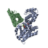
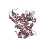
#1: Protein
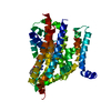
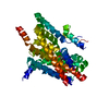
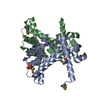
Uncharacterizedprotein
Mass: 38132.266 Da / Num. of mol.: 1 Source method: isolated from a genetically manipulated source Source: (gene. exp.) Cytophaga hutchinsonii ATCC 33406 (bacteria) Species: Cytophaga hutchinsonii / Strain: NCIMB 9469 / Gene: YP_676785.1, CHU_0153 / Plasmid: speedET / Production host: Escherichia coli (E. coli) / Strain (production host): HK100 / References: UniProt: Q11YS1, UniProt: A0A6N4SMF4*PLUS
Monochromator: Single crystal Si(111) bent (horizontal focusing) Protocol: MAD / Monochromatic (M) / Laue (L): M / Scattering type: x-ray
Radiation wavelength
ID
Wavelength (Å)
Relative weight
1
0.91837
1
2
0.97929
1
3
0.97905
1
Reflection
Resolution: 2→29.013 Å / Num. obs: 28915 / % possible obs: 99.3 % / Redundancy: 7.35 % / Biso Wilson estimate: 34.26 Å2 / Rmerge(I) obs: 0.049 / Net I/σ(I): 13.75
Reflection shell
Resolution (Å)
Rmerge(I) obs
Mean I/σ(I) obs
Num. measured obs
Diffraction-ID
% possible all
2-2.07
0.519
2.57
19575
1
95.4
2.07-2.15
0.38
3.5
20943
1
100
2.15-2.25
0.266
5
22033
1
99.9
2.25-2.37
0.198
6.5
21868
1
100
2.37-2.52
0.15
8.5
21915
1
100
2.52-2.71
0.104
11.8
21176
1
100
2.71-2.99
0.074
15.8
22151
1
100
2.99-3.42
0.049
22.1
21246
1
99.9
3.42-4.3
0.034
29.1
21149
1
100
4.3-29.013
0.027
32.4
20538
1
98.2
-
Phasing
Phasing
Method: MAD
-
Processing
Software
Name
Version
Classification
NB
REFMAC
5.2.0019
refinement
PHENIX
refinement
SHELX
phasing
MolProbity
3beta29
modelbuilding
XSCALE
datascaling
PDB_EXTRACT
2
dataextraction
MAR345
CCD
datacollection
XDS
datareduction
SHELXD
phasing
autoSHARP
phasing
Refinement
Method to determine structure: MAD / Resolution: 2→29.013 Å / Cor.coef. Fo:Fc: 0.96 / Cor.coef. Fo:Fc free: 0.945 / SU B: 7.78 / SU ML: 0.105 / TLS residual ADP flag: LIKELY RESIDUAL / Cross valid method: THROUGHOUT / σ(F): 0 / ESU R: 0.16 / ESU R Free: 0.145 Stereochemistry target values: MAXIMUM LIKELIHOOD WITH PHASES Details: 1. HYDROGENS HAVE BEEN ADDED IN THE RIDING POSITIONS. 2. A MET-INHIBITION PROTOCOL WAS USED FOR SELENOMETHIONINE INCORPORATION DURING PROTEIN EXPRESSION. THE OCCUPANCY OF THE SE ATOMS IN THE ...Details: 1. HYDROGENS HAVE BEEN ADDED IN THE RIDING POSITIONS. 2. A MET-INHIBITION PROTOCOL WAS USED FOR SELENOMETHIONINE INCORPORATION DURING PROTEIN EXPRESSION. THE OCCUPANCY OF THE SE ATOMS IN THE MSE RESIDUES WAS REDUCED TO 0.75 TO ACCOUNT FOR THE REDUCED SCATTERING POWER DUE TO PARTIAL S-MET INCORPORATION. 3. ATOM RECORD CONTAINS RESIDUAL B FACTORS ONLY. 4. PG4 MOLECULES FROM THE CRYSTALLIZATION CONDITION HAVE BEEN MODELED IN THE SOLVENT STRUCTURE.
Rfactor
Num. reflection
% reflection
Selection details
Rfree
0.218
1470
5.1 %
RANDOM
Rwork
0.184
-
-
-
all
0.185
-
-
-
obs
0.185
28893
99.88 %
-
Solvent computation
Ion probe radii: 0.8 Å / Shrinkage radii: 0.8 Å / VDW probe radii: 1.2 Å / Solvent model: MASK
Displacement parameters
Biso mean: 35.333 Å2
Baniso -1
Baniso -2
Baniso -3
1-
-0.65 Å2
-0.33 Å2
0 Å2
2-
-
-0.65 Å2
0 Å2
3-
-
-
0.98 Å2
Refinement step
Cycle: LAST / Resolution: 2→29.013 Å
Protein
Nucleic acid
Ligand
Solvent
Total
Num. atoms
2585
0
99
111
2795
Refine LS restraints
Refine-ID
Type
Dev ideal
Dev ideal target
Number
X-RAY DIFFRACTION
r_bond_refined_d
0.016
0.022
2820
X-RAY DIFFRACTION
r_bond_other_d
0.004
0.02
1960
X-RAY DIFFRACTION
r_angle_refined_deg
1.702
1.993
3802
X-RAY DIFFRACTION
r_angle_other_deg
1.304
3
4812
X-RAY DIFFRACTION
r_dihedral_angle_1_deg
3.856
5
358
X-RAY DIFFRACTION
r_dihedral_angle_2_deg
36.077
24.56
125
X-RAY DIFFRACTION
r_dihedral_angle_3_deg
12.096
15
506
X-RAY DIFFRACTION
r_dihedral_angle_4_deg
15.395
15
17
X-RAY DIFFRACTION
r_chiral_restr
0.101
0.2
433
X-RAY DIFFRACTION
r_gen_planes_refined
0.006
0.02
3056
X-RAY DIFFRACTION
r_gen_planes_other
0.003
0.02
537
X-RAY DIFFRACTION
r_nbd_refined
0.184
0.3
552
X-RAY DIFFRACTION
r_nbd_other
0.14
0.3
1979
X-RAY DIFFRACTION
r_nbtor_refined
0.169
0.5
1329
X-RAY DIFFRACTION
r_nbtor_other
0.083
0.5
1405
X-RAY DIFFRACTION
r_xyhbond_nbd_refined
0.176
0.5
166
X-RAY DIFFRACTION
r_symmetry_vdw_refined
0.093
0.3
17
X-RAY DIFFRACTION
r_symmetry_vdw_other
0.169
0.3
72
X-RAY DIFFRACTION
r_symmetry_hbond_refined
0.157
0.5
19
X-RAY DIFFRACTION
r_mcbond_it
1.993
3
1757
X-RAY DIFFRACTION
r_mcbond_other
0.491
3
679
X-RAY DIFFRACTION
r_mcangle_it
3.133
5
2760
X-RAY DIFFRACTION
r_scbond_it
5.442
8
1198
X-RAY DIFFRACTION
r_scangle_it
7.653
11
1028
LS refinement shell
Resolution: 2→2.054 Å / Total num. of bins used: 20
Rfactor
Num. reflection
% reflection
Rfree
0.298
120
-
Rwork
0.222
2006
-
obs
-
2126
99.44 %
Refinement TLS params.
Method: refined / Refine-ID: X-RAY DIFFRACTION
ID
L11 (°2)
L12 (°2)
L13 (°2)
L22 (°2)
L23 (°2)
L33 (°2)
S11 (Å °)
S12 (Å °)
S13 (Å °)
S21 (Å °)
S22 (Å °)
S23 (Å °)
S31 (Å °)
S32 (Å °)
S33 (Å °)
T11 (Å2)
T12 (Å2)
T13 (Å2)
T22 (Å2)
T23 (Å2)
T33 (Å2)
Origin x (Å)
Origin y (Å)
Origin z (Å)
1
3.3437
-2.3968
-1.6321
3.4478
1.8819
2.739
0.1382
-0.0279
0.658
-0.1541
0.2339
-0.3783
-0.2626
0.1986
-0.3721
-0.126
-0.0134
0.0078
-0.0311
0.0035
0.0522
44.892
16.3107
-7.3264
2
2.9629
0.6426
-0.3735
1.7014
0.0877
0.0653
-0.0757
0.2913
0.0995
-0.1826
0.0546
0.4432
-0.0943
0.0099
0.0211
-0.0626
0
-0.0878
0.0047
0.1317
0.0584
21.5941
7.6485
-4.5163
3
1.5212
-1.9669
-0.7496
3.1545
0.4839
0.7547
0.1104
0.0693
0.1884
-0.2276
-0.0952
-0.2161
-0.0177
-0.0354
-0.0152
-0.0634
-0.0459
-0.03
-0.0216
0.0134
-0.0792
34.3618
23.289
-6.2604
4
1.3811
0.8132
-0.9941
2.6883
-0.6201
3.196
0.1408
-0.1648
0.14
0.4378
-0.056
-0.0281
-0.2621
0.1621
-0.0848
-0.0352
-0.0136
-0.0249
-0.0105
0.0172
-0.0956
41.5408
5.669
15.0568
Refinement TLS group
Refine-ID: X-RAY DIFFRACTION / Selection: ALL / Auth asym-ID: A / Label asym-ID: A
ID
Refine TLS-ID
Auth seq-ID
Label seq-ID
1
1
1 - 46
2 - 47
2
2
47 - 179
48 - 180
3
3
180 - 223
181 - 224
4
4
224 - 329
225 - 330
+
About Yorodumi
-
News
-
Feb 9, 2022. New format data for meta-information of EMDB entries
New format data for meta-information of EMDB entries
Version 3 of the EMDB header file is now the official format.
The previous official version 1.9 will be removed from the archive.
In the structure databanks used in Yorodumi, some data are registered as the other names, "COVID-19 virus" and "2019-nCoV". Here are the details of the virus and the list of structure data.
Jan 31, 2019. EMDB accession codes are about to change! (news from PDBe EMDB page)
EMDB accession codes are about to change! (news from PDBe EMDB page)
The allocation of 4 digits for EMDB accession codes will soon come to an end. Whilst these codes will remain in use, new EMDB accession codes will include an additional digit and will expand incrementally as the available range of codes is exhausted. The current 4-digit format prefixed with “EMD-” (i.e. EMD-XXXX) will advance to a 5-digit format (i.e. EMD-XXXXX), and so on. It is currently estimated that the 4-digit codes will be depleted around Spring 2019, at which point the 5-digit format will come into force.
The EM Navigator/Yorodumi systems omit the EMD- prefix.
Related info.:Q: What is EMD? / ID/Accession-code notation in Yorodumi/EM Navigator
Yorodumi is a browser for structure data from EMDB, PDB, SASBDB, etc.
This page is also the successor to EM Navigator detail page, and also detail information page/front-end page for Omokage search.
The word "yorodu" (or yorozu) is an old Japanese word meaning "ten thousand". "mi" (miru) is to see.
Related info.:EMDB / PDB / SASBDB / Comparison of 3 databanks / Yorodumi Search / Aug 31, 2016. New EM Navigator & Yorodumi / Yorodumi Papers / Jmol/JSmol / Function and homology information / Changes in new EM Navigator and Yorodumi
 Movie
Movie Controller

 Yorodumi
 Open data
 Basic information
 Components
 Keywords
 Function and homology information
 Cytophaga hutchinsonii ATCC 33406 (bacteria)
 X-RAY DIFFRACTION /
  Authors
 Citation
 Structure visualization
 Downloads & links
 Other downloads
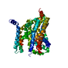


 PDBj
PDBj Assembly

 Mass: 94.971 Da / Num. of mol.: 1 / Source method: obtained synthetically / Formula: PO4
 Mass: 194.226 Da / Num. of mol.: 11 / Source method: obtained synthetically / Formula: C8H18O5 / Comment: precipitant*YM
 Mass: 18.015 Da / Num. of mol.: 111 / Source method: isolated from a natural source / Formula: H2O
 Sample preparation
 / Beamline: BL11-1 / Wavelength: 0.91837, 0.97929, 0.97905
 Processing