- PDB-2r3e: CRYSTAL STRUCTURE OF a ribokinase-like superfamily protein (EF179... -
+
Open data
ID or keywords:
Loading...
-
Basic information
Entry
Database: PDB / ID: 2r3e
Title
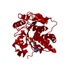
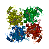
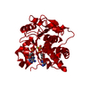
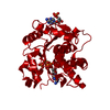
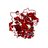
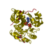
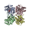
CRYSTAL STRUCTURE OF a ribokinase-like superfamily protein (EF1790) FROM ENTEROCOCCUS FAECALIS V583 AT 1.95 A RESOLUTION
Components
YjeF-related protein
Keywords
TRANSFERASE / PUTATIVE KINASE IN THE RIBOKINASE-LIKE SUPERFAMILY / STRUCTURAL GENOMICS / JOINT CENTER FOR STRUCTURAL GENOMICS / JCSG / PROTEIN STRUCTURE INITIATIVE / PSI-2
Function / homology
Function and homology information
ADP-dependent NAD(P)H-hydrate dehydratase / ADP-dependent NAD(P)H-hydrate dehydratase activity / NAD(P)HX epimerase activity / metabolite repair / nicotinamide nucleotide metabolic process / ATP binding Similarity search - Function
BIOMOLECULE: 1 SEE REMARK 350 FOR THE AUTHOR PROVIDED AND PROGRAM GENERATED ASSEMBLY INFORMATION ... BIOMOLECULE: 1 SEE REMARK 350 FOR THE AUTHOR PROVIDED AND PROGRAM GENERATED ASSEMBLY INFORMATION FOR THE STRUCTURE IN THIS ENTRY. THE REMARK MAY ALSO PROVIDE INFORMATION ON BURIED SURFACE AREA. SIZE EXCLUSION CHROMATOGRAPHY WITH STATIC LIGHT SCATTERING SUPPORTS THE ASSIGNMENT OF A TETRAMER AS A SIGNIFICANT OLIGOMERIZATION STATE.
Remark 999
SEQUENCE THE CONSTRUCT WAS EXPRESSED WITH A PURIFICATION TAG MGSDKIHHHHHHENLYFQG.
Resolution: 1.95→27.472 Å / Cor.coef. Fo:Fc: 0.974 / Cor.coef. Fo:Fc free: 0.962 / SU B: 6.15 / SU ML: 0.088 / TLS residual ADP flag: LIKELY RESIDUAL / Cross valid method: THROUGHOUT / σ(F): 0 / ESU R: 0.126 / ESU R Free: 0.122 / Stereochemistry target values: MAXIMUM LIKELIHOOD Details: 1. HYDROGENS HAVE BEEN ADDED IN THE RIDING POSITIONS. 2. A MET-INHIBITION PROTOCOL WAS USED FOR SELENOMETHIONINE INCORPORATION DURING PROTEIN EXPRESSION. THE OCCUPANCY OF THE SE ATOMS IN THE ...Details: 1. HYDROGENS HAVE BEEN ADDED IN THE RIDING POSITIONS. 2. A MET-INHIBITION PROTOCOL WAS USED FOR SELENOMETHIONINE INCORPORATION DURING PROTEIN EXPRESSION. THE OCCUPANCY OF THE SE ATOMS IN THE MSE RESIDUES WAS REDUCED TO 0.75 TO ACCOUNT FOR THE REDUCED SCATTERING POWER DUE TO PARTIAL S-MET INCORPORATION. 3. ATOM RECORD CONTAINS RESIDUAL B FACTORS ONLY. 4. ONE EDO MODELED IS PRESENT IN CRYO CONDITION.
Rfactor
Num. reflection
% reflection
Selection details
Rfree
0.194
1216
5.1 %
RANDOM
Rwork
0.155
-
-
-
all
0.157
-
-
-
obs
0.157
23697
99.84 %
-
Solvent computation
Ion probe radii: 0.8 Å / Shrinkage radii: 0.8 Å / VDW probe radii: 1.2 Å / Solvent model: BABINET MODEL WITH MASK
Displacement parameters
Biso mean: 37.69 Å2
Baniso -1
Baniso -2
Baniso -3
1-
-0.74 Å2
0 Å2
0 Å2
2-
-
-0.74 Å2
0 Å2
3-
-
-
1.49 Å2
Refinement step
Cycle: LAST / Resolution: 1.95→27.472 Å
Protein
Nucleic acid
Ligand
Solvent
Total
Num. atoms
2095
0
8
142
2245
Refine LS restraints
Refine-ID
Type
Dev ideal
Dev ideal target
Number
X-RAY DIFFRACTION
r_bond_refined_d
0.017
0.022
2155
X-RAY DIFFRACTION
r_bond_other_d
0.001
0.02
1417
X-RAY DIFFRACTION
r_angle_refined_deg
1.427
1.959
2926
X-RAY DIFFRACTION
r_angle_other_deg
0.927
3
3492
X-RAY DIFFRACTION
r_dihedral_angle_1_deg
5.969
5
279
X-RAY DIFFRACTION
r_dihedral_angle_2_deg
39.998
24.884
86
X-RAY DIFFRACTION
r_dihedral_angle_3_deg
14.108
15
364
X-RAY DIFFRACTION
r_dihedral_angle_4_deg
11.811
15
8
X-RAY DIFFRACTION
r_chiral_restr
0.085
0.2
343
X-RAY DIFFRACTION
r_gen_planes_refined
0.005
0.02
2389
X-RAY DIFFRACTION
r_gen_planes_other
0.001
0.02
401
X-RAY DIFFRACTION
r_nbd_refined
0.218
0.2
462
X-RAY DIFFRACTION
r_nbd_other
0.184
0.2
1413
X-RAY DIFFRACTION
r_nbtor_refined
0.175
0.2
1074
X-RAY DIFFRACTION
r_nbtor_other
0.088
0.2
1054
X-RAY DIFFRACTION
r_xyhbond_nbd_refined
0.146
0.2
129
X-RAY DIFFRACTION
r_symmetry_vdw_refined
0.161
0.2
11
X-RAY DIFFRACTION
r_symmetry_vdw_other
0.243
0.2
47
X-RAY DIFFRACTION
r_symmetry_hbond_refined
0.103
0.2
13
X-RAY DIFFRACTION
r_mcbond_it
2.189
3
1497
X-RAY DIFFRACTION
r_mcbond_other
0.52
3
566
X-RAY DIFFRACTION
r_mcangle_it
2.971
5
2212
X-RAY DIFFRACTION
r_scbond_it
5.451
8
848
X-RAY DIFFRACTION
r_scangle_it
6.879
11
712
LS refinement shell
Resolution: 1.95→2.001 Å / Total num. of bins used: 20
Rfactor
Num. reflection
% reflection
Rfree
0.251
81
-
Rwork
0.198
1680
-
obs
-
1761
100 %
Refinement TLS params.
Method: refined / Origin x: 44.3406 Å / Origin y: 32.1618 Å / Origin z: 0.3724 Å
11
12
13
21
22
23
31
32
33
T
-0.0108 Å2
-0.0206 Å2
0.0115 Å2
-
-0.0532 Å2
-0.0592 Å2
-
-
-0.0068 Å2
L
0.9296 °2
0.4354 °2
-0.2326 °2
-
0.8629 °2
-0.0064 °2
-
-
0.9653 °2
S
-0.0492 Å °
0.1138 Å °
-0.2609 Å °
-0.0669 Å °
-0.0147 Å °
0.0149 Å °
0.1371 Å °
-0.0761 Å °
0.064 Å °
Refinement TLS group
Selection: ALL
+
About Yorodumi
-
News
-
Feb 9, 2022. New format data for meta-information of EMDB entries
New format data for meta-information of EMDB entries
Version 3 of the EMDB header file is now the official format.
The previous official version 1.9 will be removed from the archive.
In the structure databanks used in Yorodumi, some data are registered as the other names, "COVID-19 virus" and "2019-nCoV". Here are the details of the virus and the list of structure data.
Jan 31, 2019. EMDB accession codes are about to change! (news from PDBe EMDB page)
EMDB accession codes are about to change! (news from PDBe EMDB page)
The allocation of 4 digits for EMDB accession codes will soon come to an end. Whilst these codes will remain in use, new EMDB accession codes will include an additional digit and will expand incrementally as the available range of codes is exhausted. The current 4-digit format prefixed with “EMD-” (i.e. EMD-XXXX) will advance to a 5-digit format (i.e. EMD-XXXXX), and so on. It is currently estimated that the 4-digit codes will be depleted around Spring 2019, at which point the 5-digit format will come into force.
The EM Navigator/Yorodumi systems omit the EMD- prefix.
Related info.:Q: What is EMD? / ID/Accession-code notation in Yorodumi/EM Navigator
Yorodumi is a browser for structure data from EMDB, PDB, SASBDB, etc.
This page is also the successor to EM Navigator detail page, and also detail information page/front-end page for Omokage search.
The word "yorodu" (or yorozu) is an old Japanese word meaning "ten thousand". "mi" (miru) is to see.
Related info.:EMDB / PDB / SASBDB / Comparison of 3 databanks / Yorodumi Search / Aug 31, 2016. New EM Navigator & Yorodumi / Yorodumi Papers / Jmol/JSmol / Function and homology information / Changes in new EM Navigator and Yorodumi
 Movie
Movie Controller

 Yorodumi
 Open data
 Basic information
 Components
 Keywords
 Function and homology information
 Enterococcus faecalis (bacteria)
 X-RAY DIFFRACTION /
  Authors
 Citation
 Structure visualization
 Downloads & links
 Other downloads
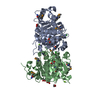
 PDBj
PDBj Assembly
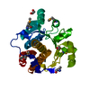

 Mass: 62.068 Da / Num. of mol.: 2 / Source method: obtained synthetically / Formula: C2H6O2
 Mass: 18.015 Da / Num. of mol.: 142 / Source method: isolated from a natural source / Formula: H2O
 Sample preparation
 / Beamline: BL11-1 / Wavelength: 1 Å
 Processing