 Movie
Movie Controller

[English] 日本語
 Yorodumi
- PDB-2qxx: Bifunctional dCTP deaminase: dUTPase from Mycobacterium tuberculo... -
+ Open data

- Basic information
| Entry | Database: PDB / ID: 2qxx | ||||||
|---|---|---|---|---|---|---|---|
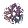
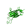
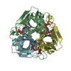
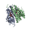
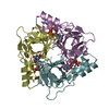
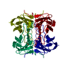
| Title | Bifunctional dCTP deaminase: dUTPase from Mycobacterium tuberculosis in complex with dTTP | ||||||
 Components Components | Deoxycytidine triphosphate deaminase | ||||||
 Keywords Keywords | HYDROLASE / distorted beta barrel / Nucleotide metabolism | ||||||
| Function / homology |  Function and homology information Function and homology informationdCTP deaminase (dUMP-forming) / dCTP deaminase (dUMP-forming) activity / dUTP biosynthetic process / dCTP deaminase activity / dUMP biosynthetic process / nucleobase-containing small molecule interconversion / dUTP diphosphatase activity / nucleotide binding Similarity search - Function | ||||||
| Biological species |   Mycobacterium tuberculosis (bacteria) Mycobacterium tuberculosis (bacteria) | ||||||
| Method |  X-RAY DIFFRACTION / SYNCHROTRON / MOLECULAR REPLACEMENT / Resolution: 2 Å X-RAY DIFFRACTION / SYNCHROTRON / MOLECULAR REPLACEMENT / Resolution: 2 Å | ||||||
 Authors Authors | Christophersen, S. / Harris, P. / Willemoes, M. | ||||||
 Citation Citation | Journal: J.Mol.Biol. / Year: 2008 Title: Mechanism of dTTP inhibition of the bifunctional dCTP deaminase:dUTPase encoded by Mycobacterium tuberculosis. Authors: Helt, S.S. / Thymark, M. / Harris, P. / Aagaard, C. / Dietrich, J. / Larsen, S. / Willemoes, M. | ||||||
| History |
|
- Structure visualization
| Structure viewer | Molecule: MolmilJmol/JSmol |
|---|
- Downloads & links
-Download
| PDBx/mmCIF format | 2qxx.cif.gz | 93.7 KB | Display | PDBx/mmCIF format |
|---|---|---|---|---|
| PDB format | pdb2qxx.ent.gz | 69.7 KB | Display | PDB format |
| PDBx/mmJSON format | 2qxx.json.gz | Tree view | PDBx/mmJSON format | |
| Others |  Other downloads Other downloads |
-Validation report
| Arichive directory | https://data.pdbj.org/pub/pdb/validation_reports/qx/2qxxftp://data.pdbj.org/pub/pdb/validation_reports/qx/2qxx | HTTPS FTP |
|---|
-Related structure data
| Related structure data |  2qlpC  1xs1S C: citing same article ( S: Starting model for refinement |
|---|---|
| Similar structure data |
-Links
 PDBj
PDBj

- Assembly
| Deposited unit | 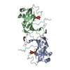
| |||||||||||||||||||||||||||||||||||||||||||||||||||||||||||||||||||||||||||||||||||||||||||
|---|---|---|---|---|---|---|---|---|---|---|---|---|---|---|---|---|---|---|---|---|---|---|---|---|---|---|---|---|---|---|---|---|---|---|---|---|---|---|---|---|---|---|---|---|---|---|---|---|---|---|---|---|---|---|---|---|---|---|---|---|---|---|---|---|---|---|---|---|---|---|---|---|---|---|---|---|---|---|---|---|---|---|---|---|---|---|---|---|---|---|---|---|
| 1 | 
| |||||||||||||||||||||||||||||||||||||||||||||||||||||||||||||||||||||||||||||||||||||||||||
| 2 | 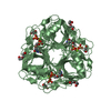
| |||||||||||||||||||||||||||||||||||||||||||||||||||||||||||||||||||||||||||||||||||||||||||
| Unit cell |
| |||||||||||||||||||||||||||||||||||||||||||||||||||||||||||||||||||||||||||||||||||||||||||
| Noncrystallographic symmetry (NCS) | NCS domain:
NCS domain segments: Ens-ID: 1 / Refine code: 4
|
-Components
| #1: Protein | Mass: 20891.604 Da / Num. of mol.: 2 Source method: isolated from a genetically manipulated source Source: (gene. exp.) Mycobacterium tuberculosis (bacteria) / Strain: H37Rv / Gene: dcd / Plasmid: pTBdcd7 / Species (production host): Escherichia coli / Production host: References: UniProt: O07247, UniProt: P9WP17*PLUS, dCTP deaminase #2: Chemical |   Mass: 24.305 Da / Num. of mol.: 2 / Source method: obtained synthetically / Formula: Mg Mass: 24.305 Da / Num. of mol.: 2 / Source method: obtained synthetically / Formula: Mg#3: Chemical |   Mass: 482.168 Da / Num. of mol.: 2 / Source method: obtained synthetically / Formula: C10H17N2O14P3 Mass: 482.168 Da / Num. of mol.: 2 / Source method: obtained synthetically / Formula: C10H17N2O14P3#4: Chemical |   Mass: 238.278 Da / Num. of mol.: 3 / Source method: obtained synthetically / Formula: C10H22O6 / Comment: precipitant*YM Mass: 238.278 Da / Num. of mol.: 3 / Source method: obtained synthetically / Formula: C10H22O6 / Comment: precipitant*YM#5: Water | ChemComp-HOH / |  Mass: 18.015 Da / Num. of mol.: 169 / Source method: isolated from a natural source / Formula: H2O Mass: 18.015 Da / Num. of mol.: 169 / Source method: isolated from a natural source / Formula: H2O |
|---|
-Experimental details
-Experiment
| Experiment | Method: X-RAY DIFFRACTION / Number of used crystals: 1 |
|---|
- Sample preparation
| Crystal | Density Matthews: 1.94 Å3/Da / Density % sol: 36.72 % |
|---|---|
| Crystal grow | Temperature: 288 K / Method: vapor diffusion, hanging drop / pH: 7.5 Details: 1.9 mg/ml enzyme in 20 mM MgCl2, 5mM dTTP, 50mM HEPES pH 6.8 reservoir solution: 45% PEG 400, 200mM MgCl2, 100 mM HEPES, pH 7.5, VAPOR DIFFUSION, HANGING DROP, temperature 288K |
-Data collection
| Diffraction | Mean temperature: 100 K |
|---|---|
| Diffraction source | Source: SYNCHROTRON / Site: ESRF  / Beamline: ID14-3 / Wavelength: 0.931 Å / Beamline: ID14-3 / Wavelength: 0.931 Å |
| Detector | Type: ADSC QUANTUM 4 / Detector: CCD / Date: Nov 17, 2006 |
| Radiation | Monochromator: Diamond (111), Ge(220) / Protocol: SINGLE WAVELENGTH / Monochromatic (M) / Laue (L): M / Scattering type: x-ray |
| Radiation wavelength | Wavelength: 0.931 Å / Relative weight: 1 |
| Reflection | Resolution: 2→19.6 Å / Num. all: 22235 / Num. obs: 22235 / % possible obs: 98.1 % / Observed criterion σ(F): -3 / Observed criterion σ(I): -3 / Biso Wilson estimate: 22.025 Å2 / Rmerge(I) obs: 0.159 / Net I/σ(I): 15.01 |
| Reflection shell | Resolution: 2→2.1 Å / Rmerge(I) obs: 0.595 / Mean I/σ(I) obs: 4.5 / Num. measured obs: 33274 / Num. unique all: 2973 / % possible all: 97.8 |
-Phasing
| Phasing MR |
|
|---|
- Processing
| Software |
| |||||||||||||||||||||||||||||||||||||||||||||||||||||||||||||||||||||||||||||||||||||||||||||||
|---|---|---|---|---|---|---|---|---|---|---|---|---|---|---|---|---|---|---|---|---|---|---|---|---|---|---|---|---|---|---|---|---|---|---|---|---|---|---|---|---|---|---|---|---|---|---|---|---|---|---|---|---|---|---|---|---|---|---|---|---|---|---|---|---|---|---|---|---|---|---|---|---|---|---|---|---|---|---|---|---|---|---|---|---|---|---|---|---|---|---|---|---|---|---|---|---|
| Refinement | Method to determine structure: MOLECULAR REPLACEMENT Starting model: PDB entry 1XS1 Resolution: 2→19.58 Å / Cor.coef. Fo:Fc: 0.959 / Cor.coef. Fo:Fc free: 0.924 / SU B: 4.257 / SU ML: 0.118 / Cross valid method: THROUGHOUT / σ(F): 0 / ESU R: 0.201 / ESU R Free: 0.171 / Stereochemistry target values: MAXIMUM LIKELIHOOD
| |||||||||||||||||||||||||||||||||||||||||||||||||||||||||||||||||||||||||||||||||||||||||||||||
| Solvent computation | Ion probe radii: 0.8 Å / Shrinkage radii: 0.8 Å / VDW probe radii: 1.4 Å / Solvent model: MASK | |||||||||||||||||||||||||||||||||||||||||||||||||||||||||||||||||||||||||||||||||||||||||||||||
| Displacement parameters | Biso mean: 16.975 Å2
| |||||||||||||||||||||||||||||||||||||||||||||||||||||||||||||||||||||||||||||||||||||||||||||||
| Refinement step | Cycle: LAST / Resolution: 2→19.58 Å
| |||||||||||||||||||||||||||||||||||||||||||||||||||||||||||||||||||||||||||||||||||||||||||||||
| Refine LS restraints |
| |||||||||||||||||||||||||||||||||||||||||||||||||||||||||||||||||||||||||||||||||||||||||||||||
| Refine LS restraints NCS | Dom-ID: 1 / Auth asym-ID: A / Ens-ID: 1 / Number: 1377 / Refine-ID: X-RAY DIFFRACTION
| |||||||||||||||||||||||||||||||||||||||||||||||||||||||||||||||||||||||||||||||||||||||||||||||
| LS refinement shell | Resolution: 2→2.043 Å / Total num. of bins used: 20
|
