 Movie
Movie Controller

[English] 日本語
 Yorodumi
- PDB-2qca: A New Crystal Form of Bovine Pancreatic RNase A in Complex with 2... -
+ Open data

- Basic information
| Entry | Database: PDB / ID: 2qca | ||||||
|---|---|---|---|---|---|---|---|
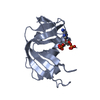
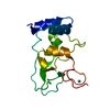
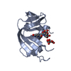
| Title | A New Crystal Form of Bovine Pancreatic RNase A in Complex with 2'-Deoxyguanosine-5'-monophosphate | ||||||
 Components Components | Ribonuclease pancreatic | ||||||
 Keywords Keywords | HYDROLASE / Ribonuclease / Protein-Nucleotide Complex / Structural Genomics / PSI-2 / Protein Structure Initiative / Center for High-Throughput Structural Biology / CHTSB | ||||||
| Function / homology |  Function and homology information Function and homology informationpancreatic ribonuclease / ribonuclease A activity / RNA nuclease activity / nucleic acid binding / defense response to Gram-positive bacterium / lyase activity / extracellular region Similarity search - Function | ||||||
| Biological species |  | ||||||
| Method |  X-RAY DIFFRACTION / MOLECULAR REPLACEMENT / Resolution: 1.33 Å X-RAY DIFFRACTION / MOLECULAR REPLACEMENT / Resolution: 1.33 Å | ||||||
 Authors Authors | Larson, S.B. / Day, J.S. / Cudney, R. / McPherson, A. / Center for High-Throughput Structural Biology (CHTSB) | ||||||
 Citation Citation | Journal: Acta Crystallogr.,Sect.F / Year: 2007 Title: A new crystal form of bovine pancreatic RNase A in complex with 2'-deoxyguanosine-5'-monophosphate. Authors: Larson, S.B. / Day, J.S. / Cudney, R. / McPherson, A. #1: Journal: Acta Crystallogr.,Sect.D / Year: 2007Title: A novel strategy for the crystallization of proteins: X-ray diffraction validation Authors: Larson, S.B. / Day, J.S. / Cudney, R. / McPherson, A. | ||||||
| History |
|
- Structure visualization
| Structure viewer | Molecule: MolmilJmol/JSmol |
|---|
- Downloads & links
-Download
| PDBx/mmCIF format | 2qca.cif.gz | 72.8 KB | Display | PDBx/mmCIF format |
|---|---|---|---|---|
| PDB format | pdb2qca.ent.gz | 54 KB | Display | PDB format |
| PDBx/mmJSON format | 2qca.json.gz | Tree view | PDBx/mmJSON format | |
| Others |  Other downloads Other downloads |
-Validation report
| Summary document | 2qca_validation.pdf.gz | 1 MB | Display | wwPDB validaton report |
|---|---|---|---|---|
| Full document | 2qca_full_validation.pdf.gz | 1 MB | Display | |
| Data in XML | 2qca_validation.xml.gz | 9.5 KB | Display | |
| Data in CIF | 2qca_validation.cif.gz | 12.8 KB | Display | |
| Arichive directory | https://data.pdbj.org/pub/pdb/validation_reports/qc/2qcaftp://data.pdbj.org/pub/pdb/validation_reports/qc/2qca | HTTPS FTP |
-Related structure data
| Related structure data | 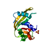 1rnoS S: Starting model for refinement |
|---|---|
| Similar structure data |


-Links
 PDBj
PDBj

- Assembly
| Deposited unit | 
| ||||||||
|---|---|---|---|---|---|---|---|---|---|
| 1 |
| ||||||||
| Unit cell |
|
-Components
| #1: Protein | Mass: 13708.326 Da / Num. of mol.: 1 / Source method: isolated from a natural source / Source: (natural) | ||||
|---|---|---|---|---|---|
| #2: Chemical |   Mass: 347.221 Da / Num. of mol.: 2 / Source method: obtained synthetically / Formula: C10H14N5O7P Mass: 347.221 Da / Num. of mol.: 2 / Source method: obtained synthetically / Formula: C10H14N5O7P#3: Water | ChemComp-HOH / |  Mass: 18.015 Da / Num. of mol.: 137 / Source method: isolated from a natural source / Formula: H2O Mass: 18.015 Da / Num. of mol.: 137 / Source method: isolated from a natural source / Formula: H2OHas protein modification | Y | |
-Experimental details
-Experiment
| Experiment | Method: X-RAY DIFFRACTION / Number of used crystals: 1 |
|---|
- Sample preparation
| Crystal | Density Matthews: 2.5 Å3/Da / Density % sol: 50.87 % |
|---|---|
| Crystal grow | Temperature: 295 K / pH: 7 Details: Reservoir: 0.6 ml of 30% PEG 3350 in 0.1 M HEPES at pH 7.0. Sample: 2 ul 10-40 mg/ml Protein in 0.1 M HEPES at pH 7.0 and 2 ul of 0.1 M DGP, cholesterol, thymine and oxamic acid in 15% PEG ...Details: Reservoir: 0.6 ml of 30% PEG 3350 in 0.1 M HEPES at pH 7.0. Sample: 2 ul 10-40 mg/ml Protein in 0.1 M HEPES at pH 7.0 and 2 ul of 0.1 M DGP, cholesterol, thymine and oxamic acid in 15% PEG 3350 and 9.1 M HEPES at pH 7.0, VAPOR DIFFUSION, SITTING DROP, temperature 295K, pH 7.00 |
-Data collection
| Diffraction | Mean temperature: 295 K |
|---|---|
| Diffraction source | Source: ROTATING ANODE / Type: RIGAKU RU200 / Wavelength: 1.5418 |
| Detector | Type: RIGAKU RAXIS / Detector: IMAGE PLATE / Date: Jan 8, 2006 / Details: OSMIC MIRRORS |
| Radiation | Monochromator: GRAPHITE / Protocol: SINGLE WAVELENGTH / Monochromatic (M) / Laue (L): M / Scattering type: x-ray |
| Radiation wavelength | Wavelength: 1.5418 Å / Relative weight: 1 |
| Reflection | Resolution: 1.33→27.54 Å / Num. obs: 27155 / % possible obs: 89.3 % / Observed criterion σ(I): 0 / Redundancy: 3.33 % / Biso Wilson estimate: 27.06 Å2 / Rmerge(I) obs: 0.049 / Net I/σ(I): 13.7 |
| Reflection shell | Resolution: 1.33→1.38 Å / Redundancy: 1.49 % / Rmerge(I) obs: 0.491 / Mean I/σ(I) obs: 0.8 / % possible all: 24.9 |
- Processing
| Software |
| |||||||||||||||||||||||||||||||||||||||||||||||||||||||||||||||||||||||||||||
|---|---|---|---|---|---|---|---|---|---|---|---|---|---|---|---|---|---|---|---|---|---|---|---|---|---|---|---|---|---|---|---|---|---|---|---|---|---|---|---|---|---|---|---|---|---|---|---|---|---|---|---|---|---|---|---|---|---|---|---|---|---|---|---|---|---|---|---|---|---|---|---|---|---|---|---|---|---|---|
| Refinement | Method to determine structure: MOLECULAR REPLACEMENT Starting model: PDB ENTRY 1RNO Resolution: 1.33→27.54 Å / Num. parameters: 10604 / Num. restraintsaints: 13217 / Cross valid method: FREE R / σ(F): 0 StereochEM target val spec case: DGP FROM PARKINSON, ET AL., ACTA CRYST. D52(1996)57-64 Stereochemistry target values: ENGH & HUBER Details: HYDROGEN ATOMS WERE INCLUDED IN RIDING POSITIONS. HYDROGEN ATOMS OF HYDROXYL GROUPS WERE PLACED BY HAND BASED ON BEST HYDROGEN BONDING INTERACTIONS. WATER MOLECULES WERE OBTAINED INITIALLY ...Details: HYDROGEN ATOMS WERE INCLUDED IN RIDING POSITIONS. HYDROGEN ATOMS OF HYDROXYL GROUPS WERE PLACED BY HAND BASED ON BEST HYDROGEN BONDING INTERACTIONS. WATER MOLECULES WERE OBTAINED INITIALLY FOUND FROM AUTOMATIC MAP INTERPRETATION AND LATER FROM MANUAL IDENTIFICATION FROM FO-FC AND 2FO-FC MAPS. WATER OCCUPANCIES WERE INITIALLY SET TO 1.0, BUT REDUCED TO 0.5 IF B VALUES EXCEEDED 80 A^2.
| |||||||||||||||||||||||||||||||||||||||||||||||||||||||||||||||||||||||||||||
| Solvent computation | Solvent model: MOEWS & KRETSINGER, J.MOL.BIOL.91(1975)201-228 | |||||||||||||||||||||||||||||||||||||||||||||||||||||||||||||||||||||||||||||
| Displacement parameters | Biso mean: 39.6 Å2 | |||||||||||||||||||||||||||||||||||||||||||||||||||||||||||||||||||||||||||||
| Refine analyze | Num. disordered residues: 12 / Occupancy sum hydrogen: 914 / Occupancy sum non hydrogen: 1105.57
| |||||||||||||||||||||||||||||||||||||||||||||||||||||||||||||||||||||||||||||
| Refinement step | Cycle: LAST / Resolution: 1.33→27.54 Å
| |||||||||||||||||||||||||||||||||||||||||||||||||||||||||||||||||||||||||||||
| Refine LS restraints |
| |||||||||||||||||||||||||||||||||||||||||||||||||||||||||||||||||||||||||||||
| LS refinement shell |
|
