 ムービー
ムービー コントローラー


+ データを開く

- 基本情報
基本情報
| 登録情報 | データベース: PDB / ID: 6qmn | ||||||
|---|---|---|---|---|---|---|---|
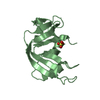
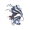
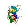
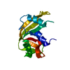
| タイトル | Crystal structure of a Ribonuclease A-Onconase chimera | ||||||
 要素 要素 | Ribonuclease pancreatic | ||||||
 キーワード キーワード | HYDROLASE / pancreatic ribonuclease A / alpha/beta fold / chimera | ||||||
| 機能・相同性 |  機能・相同性情報 機能・相同性情報pancreatic ribonuclease / ribonuclease A activity / nucleic acid binding / defense response to Gram-positive bacterium / hydrolase activity / extracellular region 類似検索 - 分子機能 | ||||||
| 生物種 |  Bison bison (アメリカバイソン) Bison bison (アメリカバイソン) | ||||||
| 手法 |  X線回折 / 分子置換 / 解像度: 2.31 Å X線回折 / 分子置換 / 解像度: 2.31 Å | ||||||
 データ登録者 データ登録者 | Esposito, L. / Vitagliano, L. / Ruggiero, A. / Picone, D. / Leone, S. / Donnarumma, F. | ||||||
 引用 引用 | ジャーナル: Int.J.Biol.Macromol. / 年: 2019 タイトル: Structure, stability and aggregation propensity of a Ribonuclease A-Onconase chimera. 著者: Esposito, L. / Donnarumma, F. / Ruggiero, A. / Leone, S. / Vitagliano, L. / Picone, D. | ||||||
| 履歴 |
|
- 構造の表示
構造の表示
| 構造ビューア | 分子: MolmilJmol/JSmol |
|---|
- ダウンロードとリンク
-ダウンロード
| PDBx/mmCIF形式 | 6qmn.cif.gz | 86 KB | 表示 | PDBx/mmCIF形式 |
|---|---|---|---|---|
| PDB形式 | pdb6qmn.ent.gz | 65.4 KB | 表示 | PDB形式 |
| PDBx/mmJSON形式 | 6qmn.json.gz | ツリー表示 | PDBx/mmJSON形式 | |
| その他 |  その他のダウンロード その他のダウンロード |
-検証レポート
| アーカイブディレクトリ | https://data.pdbj.org/pub/pdb/validation_reports/qm/6qmnftp://data.pdbj.org/pub/pdb/validation_reports/qm/6qmn | HTTPS FTP |
|---|
-関連構造データ
| 関連構造データ | 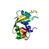 1kf4S S: 精密化の開始モデル |
|---|---|
| 類似構造データ |


-リンク
 PDBj
PDBj

- 集合体
集合体
| 登録構造単位 | 
| ||||||||
|---|---|---|---|---|---|---|---|---|---|
| 1 | 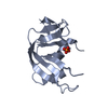
| ||||||||
| 2 | 
| ||||||||
| 3 | 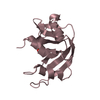
| ||||||||
| 単位格子 |
|
-要素
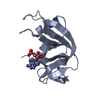
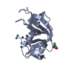
| #1: タンパク質 | 分子量: 13491.060 Da / 分子数: 3 / 由来タイプ: 組換発現 由来: (組換発現) Bison bison (アメリカバイソン)遺伝子: RNASE1, RNS1 / 発現宿主:  #2: 化合物 |   分子量: 94.971 Da / 分子数: 3 / 由来タイプ: 合成 / 式: PO4 分子量: 94.971 Da / 分子数: 3 / 由来タイプ: 合成 / 式: PO4#3: 水 | ChemComp-HOH / |  分子量: 18.015 Da / 分子数: 154 / 由来タイプ: 天然 / 式: H2O 分子量: 18.015 Da / 分子数: 154 / 由来タイプ: 天然 / 式: H2OHas protein modification | Y | |
|---|
-実験情報
-実験
| 実験 | 手法: X線回折 / 使用した結晶の数: 1 |
|---|
- 試料調製
試料調製
| 結晶 | マシュー密度: 2.47 Å3/Da / 溶媒含有率: 50.17 % |
|---|---|
| 結晶化 | 温度: 296 K / 手法: 蒸気拡散法, ハンギングドロップ法 詳細: 0.100 M Bis-Tris pH 6.5, 28 % W/V Polyethylene glycol monomethyl ether 2000 |
-データ収集
| 回折 | 平均測定温度: 100 K / Serial crystal experiment: N |
|---|---|
| 放射光源 | 由来: 回転陽極 / タイプ: RIGAKU MICROMAX-007 / 波長: 1.5419 Å |
| 検出器 | タイプ: RIGAKU SATURN 944 / 検出器: CCD / 日付: 2015年10月30日 |
| 放射 | プロトコル: SINGLE WAVELENGTH / 単色(M)・ラウエ(L): M / 散乱光タイプ: x-ray |
| 放射波長 | 波長: 1.5419 Å / 相対比: 1 |
| 反射 | 解像度: 2.31→28 Å / Num. obs: 16885 / % possible obs: 96.3 % / 冗長度: 2.8 % / Rmerge(I) obs: 0.126 / Net I/σ(I): 10.2 |
| 反射 シェル | 解像度: 2.31→2.38 Å / 冗長度: 2.7 % / Rmerge(I) obs: 0.368 / Mean I/σ(I) obs: 2.7 / Num. unique obs: 1664 / % possible all: 96.1 |
- 解析
解析
| ソフトウェア |
| ||||||||||||||||||||||||||||||||||||||||||||||||||||||||||||
|---|---|---|---|---|---|---|---|---|---|---|---|---|---|---|---|---|---|---|---|---|---|---|---|---|---|---|---|---|---|---|---|---|---|---|---|---|---|---|---|---|---|---|---|---|---|---|---|---|---|---|---|---|---|---|---|---|---|---|---|---|---|
| 精密化 | 構造決定の手法: 分子置換 開始モデル: 1kf4 解像度: 2.31→27.85 Å / Cor.coef. Fo:Fc: 0.936 / Cor.coef. Fo:Fc free: 0.871 / SU B: 12.029 / SU ML: 0.281 / 交差検証法: THROUGHOUT / σ(F): 0 / ESU R: 0.421 / ESU R Free: 0.303 詳細: HYDROGENS HAVE BEEN ADDED IN THE RIDING POSITIONS U VALUES : REFINED INDIVIDUALLY
| ||||||||||||||||||||||||||||||||||||||||||||||||||||||||||||
| 溶媒の処理 | イオンプローブ半径: 0.8 Å / 減衰半径: 0.8 Å / VDWプローブ半径: 1.2 Å | ||||||||||||||||||||||||||||||||||||||||||||||||||||||||||||
| 原子変位パラメータ | Biso max: 106.21 Å2 / Biso mean: 46.698 Å2 / Biso min: 23.81 Å2
| ||||||||||||||||||||||||||||||||||||||||||||||||||||||||||||
| 精密化ステップ | サイクル: final / 解像度: 2.31→27.85 Å
| ||||||||||||||||||||||||||||||||||||||||||||||||||||||||||||
| 拘束条件 |
| ||||||||||||||||||||||||||||||||||||||||||||||||||||||||||||
| LS精密化 シェル | 解像度: 2.309→2.369 Å / Rfactor Rfree error: 0 / Total num. of bins used: 20
|
