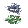
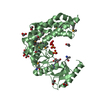
BIOMOLECULE: 1,2 THIS ENTRY CONTAINS THE CRYSTALLOGRAPHIC ASYMMETRIC UNIT WHICH CONSISTS OF 2 ... BIOMOLECULE: 1,2 THIS ENTRY CONTAINS THE CRYSTALLOGRAPHIC ASYMMETRIC UNIT WHICH CONSISTS OF 2 CHAIN(S). SEE REMARK 350 FOR INFORMATION ON GENERATING THE BIOLOGICAL MOLECULE(S). SIZE EXCLUSION CHROMATOGRAPHY SUPPORTS THE ASSIGNMENT OF A MONOMER AS A SIGNIFICANT OLIGOMERIZATION STATE.
Remark 999
SEQUENCE THE CONSTRUCT WAS EXPRESSED WITH AN N-TERMINAL PURIFICATION TAG MGSDKIHHHHHHENLYFQG. THE ... SEQUENCE THE CONSTRUCT WAS EXPRESSED WITH AN N-TERMINAL PURIFICATION TAG MGSDKIHHHHHHENLYFQG. THE TAG WAS REMOVED WITH TEV PROTEASE LEAVING ONLY A GLYCINE FOLLOWED BY THE TARGET SEQUENCE.
Type: MARMOSAIC 325 mm CCD / Detector: CCD / Date: May 4, 2007 / Details: Flat mirror (vertical focusing)
Radiation
Monochromator: Single crystal Si(111) bent (horizontal focusing) Protocol: MAD / Monochromatic (M) / Laue (L): M / Scattering type: x-ray
Radiation wavelength
ID
Wavelength (Å)
Relative weight
1
0.91837
1
2
0.97904
1
3
0.97932
1
Reflection
Resolution: 2.5→29.961 Å / Num. obs: 59155 / % possible obs: 99.9 % / Redundancy: 10.8 % / Biso Wilson estimate: 50.53 Å2 / Rmerge(I) obs: 0.126 / Rsym value: 0.126 / Net I/σ(I): 4.6
Reflection shell
Diffraction-ID: 1
Resolution (Å)
Redundancy (%)
Rmerge(I) obs
Mean I/σ(I) obs
Num. measured all
Num. unique all
Rsym value
% possible all
2.5-2.56
11
0.864
0.9
47319
4302
0.864
100
2.56-2.64
11
0.676
1.1
46118
4195
0.676
100
2.64-2.71
11
0.599
1.2
44831
4087
0.599
100
2.71-2.8
11
0.463
1.6
43474
3961
0.463
100
2.8-2.89
11
0.385
1.9
42498
3870
0.385
100
2.89-2.99
11
0.308
2.4
40839
3716
0.308
100
2.99-3.1
11
0.247
3
39532
3609
0.247
100
3.1-3.23
10.9
0.198
3.8
38079
3480
0.198
100
3.23-3.37
10.9
0.149
4.9
36487
3338
0.149
100
3.37-3.54
10.9
0.123
5.6
34768
3202
0.123
100
3.54-3.73
10.9
0.096
3.2
33111
3047
0.096
100
3.73-3.95
10.8
0.085
4.6
31302
2904
0.085
100
3.95-4.23
10.8
0.075
8.5
29469
2737
0.075
100
4.23-4.56
10.7
0.077
8.4
27343
2563
0.077
100
4.56-5
10.6
0.077
8
24873
2351
0.077
100
5-5.59
10.5
0.071
8.8
22795
2162
0.071
100
5.59-6.45
10.5
0.077
8.4
20114
1922
0.077
100
6.45-7.91
10.2
0.069
9.3
16927
1654
0.069
100
7.91-11.18
9.9
0.048
12.2
12959
1310
0.048
100
11.18-29.96
8.9
0.051
10.5
6665
745
0.051
94.8
-
Phasing
Phasing
Method: MAD
-
Processing
Software
Name
Version
Classification
NB
REFMAC
5.2.0019
refinement
PHENIX
refinement
MolProbity
3beta29
modelbuilding
SCALA
datascaling
PDB_EXTRACT
3
dataextraction
MAR345
CCD
datacollection
MOSFLM
datareduction
SOLVE
phasing
RESOLVE
phasing
Refinement
Method to determine structure: MAD / Resolution: 2.5→29.961 Å / Cor.coef. Fo:Fc: 0.941 / Cor.coef. Fo:Fc free: 0.934 / SU B: 10.512 / SU ML: 0.119 / TLS residual ADP flag: LIKELY RESIDUAL / Cross valid method: THROUGHOUT / σ(F): 0 / ESU R: 0.206 / ESU R Free: 0.169 Stereochemistry target values: MAXIMUM LIKELIHOOD WITH PHASES Details: 1. HYDROGENS HAVE BEEN ADDED IN THE RIDING POSITIONS. 2. ATOM RECORD CONTAINS RESIDUAL B FACTORS ONLY. 3. A MET-INHIBITION PROTOCOL WAS USED FOR SELENOMETHIONINE INCORPORATION DURING PROTEIN ...Details: 1. HYDROGENS HAVE BEEN ADDED IN THE RIDING POSITIONS. 2. ATOM RECORD CONTAINS RESIDUAL B FACTORS ONLY. 3. A MET-INHIBITION PROTOCOL WAS USED FOR SELENOMETHIONINE INCORPORATION DURING PROTEIN EXPRESSION. THE OCCUPANCY OF THE SE ATOMS IN THE MSE RESIDUES WAS REDUCED TO 0.75 FOR THE REDUCED SCATTERING POWER DUE TO PARTIAL S-MET INCORPORATION. 4. ELECTRON DENSITIES CORRESPONDING TO RESIDUES 1-21 AND 53-56 IN THE A SUBUNIT AND RESIDUES 1-21 AND 52-55 IN THE B SUBUNIT WERE DISORDERED. THEREFORE, THESE RESIDUES WERE NOT MODELED. 5. CITRIC ACID AND SULFATE MOLECULES FROM THE CRYSTALLIZATION SOLUTION WERE MODELED INTO THE STRUCTURE. THE OCCUPANCIES OF THE SULFATES WERE REDUCED TO ACCOUNT FOR THE REDUCED SCATTERING. 6. SEVERAL MOLECULES OF ETHYLENE GLYCOL, USED AS A CRYOPROTECTANT, WERE MODELED INTO THE STRUCTURE. 7. ADENOSINE WAS MODELED INTO EACH SUBUNIT. THE POSITIONING OF THIS LIGAND IS BASED ON A SIMILAR POSITIONING OF AN ADENOSINE MOIETY IN RELATED STRUCTURES. 8. AN UNKNOWN LIGAND (UNL) WAS MODELED INTO EACH SUBUNIT. 9. ALA A24 AND ASP A65 ARE IN POOR REGIONS OF ELECTRON DENSITY AND ARE RAMACHANDRAN OUTLIERS.
Rfactor
Num. reflection
% reflection
Selection details
Rfree
0.21
2988
5.1 %
RANDOM
Rwork
0.198
-
-
-
obs
0.199
59108
99.91 %
-
Solvent computation
Ion probe radii: 0.8 Å / Shrinkage radii: 0.8 Å / VDW probe radii: 1.4 Å / Solvent model: BABINET MODEL WITH MASK
In the structure databanks used in Yorodumi, some data are registered as the other names, "COVID-19 virus" and "2019-nCoV". Here are the details of the virus and the list of structure data.
Jan 31, 2019. EMDB accession codes are about to change! (news from PDBe EMDB page)
EMDB accession codes are about to change! (news from PDBe EMDB page)
The allocation of 4 digits for EMDB accession codes will soon come to an end. Whilst these codes will remain in use, new EMDB accession codes will include an additional digit and will expand incrementally as the available range of codes is exhausted. The current 4-digit format prefixed with “EMD-” (i.e. EMD-XXXX) will advance to a 5-digit format (i.e. EMD-XXXXX), and so on. It is currently estimated that the 4-digit codes will be depleted around Spring 2019, at which point the 5-digit format will come into force.
The EM Navigator/Yorodumi systems omit the EMD- prefix.
Related info.:Q: What is EMD? / ID/Accession-code notation in Yorodumi/EM Navigator
Yorodumi is a browser for structure data from EMDB, PDB, SASBDB, etc.
This page is also the successor to EM Navigator detail page, and also detail information page/front-end page for Omokage search.
The word "yorodu" (or yorozu) is an old Japanese word meaning "ten thousand". "mi" (miru) is to see.
Related info.:EMDB / PDB / SASBDB / Comparison of 3 databanks / Yorodumi Search / Aug 31, 2016. New EM Navigator & Yorodumi / Yorodumi Papers / Jmol/JSmol / Function and homology information / Changes in new EM Navigator and Yorodumi
 Movie
Movie Controller

 Yorodumi
 Open data
 Basic information
 Components
 Keywords
 Function and homology information
 X-RAY DIFFRACTION /
  Authors
 Citation
 Structure visualization
 Downloads & links
 Other downloads
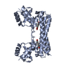


 PDBj
PDBj


 Assembly
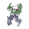


 Mass: 267.241 Da / Num. of mol.: 2 / Source method: obtained synthetically / Formula: C10H13N5O4
 Mass: 62.068 Da / Num. of mol.: 8 / Source method: obtained synthetically / Formula: C2H6O2
 Mass: 96.063 Da / Num. of mol.: 2 / Source method: obtained synthetically / Formula: SO4
 Mass: 192.124 Da / Num. of mol.: 1 / Source method: obtained synthetically / Formula: C6H8O7
 Sample preparation
 / Beamline: BL11-1 / Wavelength: 0.91837, 0.97904, 0.97932
 Processing