 Movie
Movie Controller


+ Open data

- Basic information
| Entry | Database: PDB / ID: 2q5f | ||||||
|---|---|---|---|---|---|---|---|
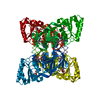
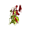
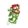
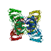
| Title | Crystal structure of LMNADK1 from Listeria monocytogenes | ||||||
 Components Components | Probable inorganic polyphosphate/ATP-NAD kinase 1 | ||||||
 Keywords Keywords | TRANSFERASE / Mutant | ||||||
| Function / homology |  Function and homology information Function and homology informationNAD+ kinase / NAD+ kinase activity / NADP+ biosynthetic process / NAD+ metabolic process / NAD binding / ATP binding / metal ion binding / cytoplasm Similarity search - Function | ||||||
| Biological species |  Listeria monocytogenes EGD-e (bacteria) Listeria monocytogenes EGD-e (bacteria) | ||||||
| Method |  X-RAY DIFFRACTION / SYNCHROTRON / MOLECULAR REPLACEMENT / Resolution: 1.9 Å X-RAY DIFFRACTION / SYNCHROTRON / MOLECULAR REPLACEMENT / Resolution: 1.9 Å | ||||||
 Authors Authors | Poncet-Montange, G. / Assairi, L. / Arold, S. / Pochet, S. / Labesse, G. | ||||||
 Citation Citation | Journal: J.Biol.Chem. / Year: 2007 Title: NAD kinases use substrate-assisted catalysis for specific recognition of NAD. Authors: Poncet-Montange, G. / Assairi, L. / Arold, S. / Pochet, S. / Labesse, G. | ||||||
| History |
|
- Structure visualization
| Structure viewer | Molecule: MolmilJmol/JSmol |
|---|
- Downloads & links
-Download
| PDBx/mmCIF format | 2q5f.cif.gz | 68.7 KB | Display | PDBx/mmCIF format |
|---|---|---|---|---|
| PDB format | pdb2q5f.ent.gz | 50.8 KB | Display | PDB format |
| PDBx/mmJSON format | 2q5f.json.gz | Tree view | PDBx/mmJSON format | |
| Others |  Other downloads Other downloads |
-Validation report
| Arichive directory | https://data.pdbj.org/pub/pdb/validation_reports/q5/2q5fftp://data.pdbj.org/pub/pdb/validation_reports/q5/2q5f | HTTPS FTP |
|---|
-Related structure data
| Related structure data |  2i1wC 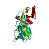 2i29C 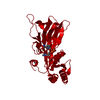 2i2aC  2i2bC  2i2cC 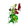 2i2dC 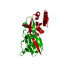 2i2fC  4dy6C C: citing same article ( |
|---|---|
| Similar structure data |


-Links
 PDBj
PDBj- Assembly
| Deposited unit | 
| ||||||||
|---|---|---|---|---|---|---|---|---|---|
| 1 | 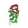
| ||||||||
| Unit cell |
|
-Components
| #1: Protein | Mass: 31036.244 Da / Num. of mol.: 1 / Mutation: H223E Source method: isolated from a genetically manipulated source Source: (gene. exp.) Listeria monocytogenes EGD-e (bacteria)Species: Listeria monocytogenes / Strain: EGD-E / Gene: ppnK1 / Plasmid: PET22B / Species (production host): Escherichia coli / Production host: |
|---|---|
| #2: Chemical | ChemComp-DTA / (  Mass: 564.598 Da / Num. of mol.: 1 / Source method: obtained synthetically / Formula: C20H24N10O6S2 Mass: 564.598 Da / Num. of mol.: 1 / Source method: obtained synthetically / Formula: C20H24N10O6S2 |
| #3: Water | ChemComp-HOH /  Mass: 18.015 Da / Num. of mol.: 84 / Source method: isolated from a natural source / Formula: H2O Mass: 18.015 Da / Num. of mol.: 84 / Source method: isolated from a natural source / Formula: H2O |
-Experimental details
-Experiment
| Experiment | Method: X-RAY DIFFRACTION / Number of used crystals: 1 |
|---|
- Sample preparation
| Crystal | Density Matthews: 2.26 Å3/Da / Density % sol: 45.46 % |
|---|---|
| Crystal grow | Temperature: 291 K / Method: vapor diffusion, hanging drop / pH: 5.5 Details: PEG400 15-20% w/v, KCitrate 50 mM, KCl 300 mM, pH 5.5, VAPOR DIFFUSION, HANGING DROP, temperature 291K |
-Data collection
| Diffraction | Mean temperature: 100 K |
|---|---|
| Diffraction source | Source: SYNCHROTRON / Site: ESRF  / Beamline: ID14-2 / Wavelength: 0.933 / Beamline: ID14-2 / Wavelength: 0.933 |
| Detector | Type: ADSC QUANTUM 210 / Detector: CCD / Date: Nov 13, 2006 |
| Radiation | Protocol: SINGLE WAVELENGTH / Monochromatic (M) / Laue (L): M / Scattering type: x-ray |
| Radiation wavelength | Wavelength: 0.933 Å / Relative weight: 1 |
| Reflection | Resolution: 1.9→28.2 Å / Num. all: 22000 / Num. obs: 19663 / % possible obs: 87.5 % / Observed criterion σ(F): 0 / Observed criterion σ(I): 0 / Redundancy: 5.9 % / Biso Wilson estimate: 29.25 Å2 / Rmerge(I) obs: 0.06 / Rsym value: 0.063 / Net I/σ(I): 20.2 |
| Reflection shell | Resolution: 1.9→1.99 Å / Redundancy: 3.9 % / Rmerge(I) obs: 0.489 / Mean I/σ(I) obs: 2.5 / Num. unique all: 1644 / % possible all: 51.6 |
- Processing
| Software |
| |||||||||||||||||||||||||||||||||||||||||||||||||||||||||||||||||||||||||||||||||||||||||||||||||||||||||||||||||||||||||||||||||||||||||||||||||||||||||||||||||||||||||||||||
|---|---|---|---|---|---|---|---|---|---|---|---|---|---|---|---|---|---|---|---|---|---|---|---|---|---|---|---|---|---|---|---|---|---|---|---|---|---|---|---|---|---|---|---|---|---|---|---|---|---|---|---|---|---|---|---|---|---|---|---|---|---|---|---|---|---|---|---|---|---|---|---|---|---|---|---|---|---|---|---|---|---|---|---|---|---|---|---|---|---|---|---|---|---|---|---|---|---|---|---|---|---|---|---|---|---|---|---|---|---|---|---|---|---|---|---|---|---|---|---|---|---|---|---|---|---|---|---|---|---|---|---|---|---|---|---|---|---|---|---|---|---|---|---|---|---|---|---|---|---|---|---|---|---|---|---|---|---|---|---|---|---|---|---|---|---|---|---|---|---|---|---|---|---|---|---|---|
| Refinement | Method to determine structure: MOLECULAR REPLACEMENT Starting model: PDB2I2C Resolution: 1.9→28.2 Å / Cor.coef. Fo:Fc: 0.956 / Cor.coef. Fo:Fc free: 0.95 / SU B: 8.046 / SU ML: 0.106 / TLS residual ADP flag: LIKELY RESIDUAL / Cross valid method: THROUGHOUT / σ(F): 0 / σ(I): 0 / ESU R: 0.175 / ESU R Free: 0.152 / Stereochemistry target values: MAXIMUM LIKELIHOOD / Details: HYDROGENS HAVE BEEN ADDED IN THE RIDING POSITIONS
| |||||||||||||||||||||||||||||||||||||||||||||||||||||||||||||||||||||||||||||||||||||||||||||||||||||||||||||||||||||||||||||||||||||||||||||||||||||||||||||||||||||||||||||||
| Solvent computation | Ion probe radii: 0.8 Å / Shrinkage radii: 0.8 Å / VDW probe radii: 1.4 Å / Solvent model: MASK | |||||||||||||||||||||||||||||||||||||||||||||||||||||||||||||||||||||||||||||||||||||||||||||||||||||||||||||||||||||||||||||||||||||||||||||||||||||||||||||||||||||||||||||||
| Displacement parameters | Biso mean: 27.827 Å2
| |||||||||||||||||||||||||||||||||||||||||||||||||||||||||||||||||||||||||||||||||||||||||||||||||||||||||||||||||||||||||||||||||||||||||||||||||||||||||||||||||||||||||||||||
| Refine analyze | Luzzati coordinate error obs: 0.3036 Å | |||||||||||||||||||||||||||||||||||||||||||||||||||||||||||||||||||||||||||||||||||||||||||||||||||||||||||||||||||||||||||||||||||||||||||||||||||||||||||||||||||||||||||||||
| Refinement step | Cycle: LAST / Resolution: 1.9→28.2 Å
| |||||||||||||||||||||||||||||||||||||||||||||||||||||||||||||||||||||||||||||||||||||||||||||||||||||||||||||||||||||||||||||||||||||||||||||||||||||||||||||||||||||||||||||||
| Refine LS restraints |
| |||||||||||||||||||||||||||||||||||||||||||||||||||||||||||||||||||||||||||||||||||||||||||||||||||||||||||||||||||||||||||||||||||||||||||||||||||||||||||||||||||||||||||||||
| LS refinement shell | Resolution: 1.9→1.949 Å / Total num. of bins used: 20
| |||||||||||||||||||||||||||||||||||||||||||||||||||||||||||||||||||||||||||||||||||||||||||||||||||||||||||||||||||||||||||||||||||||||||||||||||||||||||||||||||||||||||||||||
| Refinement TLS params. | Method: refined / Refine-ID: X-RAY DIFFRACTION
| |||||||||||||||||||||||||||||||||||||||||||||||||||||||||||||||||||||||||||||||||||||||||||||||||||||||||||||||||||||||||||||||||||||||||||||||||||||||||||||||||||||||||||||||
| Refinement TLS group |
|
