 Movie
Movie Controller

[English] 日本語
 Yorodumi
- PDB-2q22: Crystal structure of uncharacterized protein (YP_323524.1) from A... -
+ Open data

- Basic information
| Entry | Database: PDB / ID: 2q22 | ||||||
|---|---|---|---|---|---|---|---|
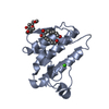
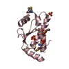
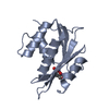
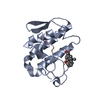
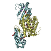
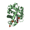
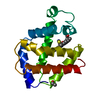
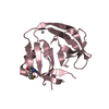
| Title | Crystal structure of uncharacterized protein (YP_323524.1) from Anabaena variabilis ATCC 29413 at 2.11 A resolution | ||||||
 Components Components | Uncharacterized protein | ||||||
 Keywords Keywords | STRUCTURAL GENOMICS / UNKNOWN FUNCTION / YP_323524.1 / uncharacterized protein / Joint Center for Structural Genomics / JCSG / Protein Structure Initiative / PSI-2 | ||||||
| Function / homology | Protein of unknown function DUF1824 / Domain of unknown function (DUF1824) / Dihydrodipicolinate Reductase; domain 2 / Dihydrodipicolinate Reductase; domain 2 / 2-Layer Sandwich / Alpha Beta / ACETATE ION / DUF1824 domain-containing protein Function and homology information Function and homology information | ||||||
| Biological species |  Anabaena variabilis ATCC 29413 (bacteria) Anabaena variabilis ATCC 29413 (bacteria) | ||||||
| Method |  X-RAY DIFFRACTION / SYNCHROTRON / MAD / Resolution: 2.11 Å X-RAY DIFFRACTION / SYNCHROTRON / MAD / Resolution: 2.11 Å | ||||||
 Authors Authors | Joint Center for Structural Genomics (JCSG) | ||||||
 Citation Citation | Journal: To be published Title: Crystal structure of uncharacterized protein (YP_323524.1) from Anabaena variabilis ATCC 29413 at 2.11 A resolution Authors: Joint Center for Structural Genomics (JCSG) | ||||||
| History |
| ||||||
| Remark 300 | THIS ENTRY CONTAINS THE CRYSTALLOGRAPHIC ASYMMETRIC UNIT WHICH CONSISTS OF 3 CHAIN(S). SEE REMARK ... THIS ENTRY CONTAINS THE CRYSTALLOGRAPHIC ASYMMETRIC UNIT WHICH CONSISTS OF 3 CHAIN(S). SEE REMARK 350 FOR INFORMATION ON GENERATING THE BIOLOGICAL MOLECULE(S). SIZE EXCLUSION CHROMATOGRAPHY SUPPORTS THE ASSIGNMENT OF A MONOMER AS A OLIGOMERIZATION STATE IN SOLUTION. | ||||||
| Remark 999 | SEQUENCE THE CONSTRUCT WAS EXPRESSED WITH A PURIFICATION TAG MGSDKIHHHHHHENLYFQG. THE TAG WAS ... SEQUENCE THE CONSTRUCT WAS EXPRESSED WITH A PURIFICATION TAG MGSDKIHHHHHHENLYFQG. THE TAG WAS REMOVED WITH TEV PROTEASE LEAVING ONLY A GLYCINE FOLLOWED BY THE TARGET SEQUENCE. |
- Structure visualization
| Structure viewer | Molecule: MolmilJmol/JSmol |
|---|
- Downloads & links
-Download
| PDBx/mmCIF format | 2q22.cif.gz | 95.1 KB | Display | PDBx/mmCIF format |
|---|---|---|---|---|
| PDB format | pdb2q22.ent.gz | 72 KB | Display | PDB format |
| PDBx/mmJSON format | 2q22.json.gz | Tree view | PDBx/mmJSON format | |
| Others |  Other downloads Other downloads |
-Validation report
| Arichive directory | https://data.pdbj.org/pub/pdb/validation_reports/q2/2q22ftp://data.pdbj.org/pub/pdb/validation_reports/q2/2q22 | HTTPS FTP |
|---|
-Related structure data
| Similar structure data | |
|---|---|
| Other databases |


-Links
 PDBj
PDBj- Assembly
| Deposited unit | 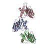
| ||||||||||||||||||||||||
|---|---|---|---|---|---|---|---|---|---|---|---|---|---|---|---|---|---|---|---|---|---|---|---|---|---|
| 1 | 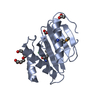
| ||||||||||||||||||||||||
| 2 | 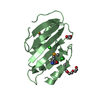
| ||||||||||||||||||||||||
| 3 | 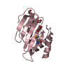
| ||||||||||||||||||||||||
| Unit cell |
| ||||||||||||||||||||||||
| Noncrystallographic symmetry (NCS) | NCS domain:
NCS domain segments: Component-ID: 1 / Ens-ID: 1 / Beg auth comp-ID: LEU / Beg label comp-ID: LEU / End auth comp-ID: VAL / End label comp-ID: VAL / Refine code: 2 / Auth seq-ID: 9 - 138 / Label seq-ID: 10 - 139
| ||||||||||||||||||||||||
| Details | SIZE EXCLUSION CHROMATOGRAPHY SUPPORTS THE ASSIGNMENT OF A MONOMER AS A OLIGOMERIZATION STATE IN SOLUTION. |
-Components
-Protein , 1 types, 3 molecules ABC
| #1: Protein | Mass: 15488.285 Da / Num. of mol.: 3 Source method: isolated from a genetically manipulated source Source: (gene. exp.) Anabaena variabilis ATCC 29413 (bacteria)Species: Anabaena variabilis / Strain: PCC 7937 / Gene: YP_323524.1, Ava_3019 / Plasmid: speedET / Production host: |
|---|
-Non-polymers , 5 types, 202 molecules 


| #2: Chemical | ChemComp-ACT /  Mass: 59.044 Da / Num. of mol.: 4 / Source method: obtained synthetically / Formula: C2H3O2 Mass: 59.044 Da / Num. of mol.: 4 / Source method: obtained synthetically / Formula: C2H3O2#3: Chemical |  Mass: 35.453 Da / Num. of mol.: 3 / Source method: obtained synthetically / Formula: Cl Mass: 35.453 Da / Num. of mol.: 3 / Source method: obtained synthetically / Formula: Cl#4: Chemical |  Mass: 194.226 Da / Num. of mol.: 2 / Source method: obtained synthetically / Formula: C8H18O5 / Comment: precipitant*YM Mass: 194.226 Da / Num. of mol.: 2 / Source method: obtained synthetically / Formula: C8H18O5 / Comment: precipitant*YM#5: Chemical |  Mass: 62.068 Da / Num. of mol.: 2 / Source method: obtained synthetically / Formula: C2H6O2 Mass: 62.068 Da / Num. of mol.: 2 / Source method: obtained synthetically / Formula: C2H6O2#6: Water | ChemComp-HOH / | Mass: 18.015 Da / Num. of mol.: 191 / Source method: isolated from a natural source / Formula: H2O |
|---|
-Details
| Has protein modification | Y |
|---|
-Experimental details
-Experiment
| Experiment | Method: X-RAY DIFFRACTION / Number of used crystals: 1 |
|---|
- Sample preparation
| Crystal | Density Matthews: 2.84 Å3/Da / Density % sol: 56.75 % |
|---|---|
| Crystal grow | Temperature: 293 K / Method: vapor diffusion, sitting drop / pH: 4.18 Details: NANODROP, 5.0% PEG 1000, 48.3% Ethylene glycol, 0.1M Sodium acetate pH 4.18, VAPOR DIFFUSION, SITTING DROP, temperature 293K |
-Data collection
| Diffraction | Mean temperature: 100 K | ||||||||||||||||||||||||||||||||||||||||||||||||||||||||||||||||||||||||||||||||||||||||||||||||||||||||||||||||||||||||||||||||||||||||||||||||||||||||||||||||||||||||
|---|---|---|---|---|---|---|---|---|---|---|---|---|---|---|---|---|---|---|---|---|---|---|---|---|---|---|---|---|---|---|---|---|---|---|---|---|---|---|---|---|---|---|---|---|---|---|---|---|---|---|---|---|---|---|---|---|---|---|---|---|---|---|---|---|---|---|---|---|---|---|---|---|---|---|---|---|---|---|---|---|---|---|---|---|---|---|---|---|---|---|---|---|---|---|---|---|---|---|---|---|---|---|---|---|---|---|---|---|---|---|---|---|---|---|---|---|---|---|---|---|---|---|---|---|---|---|---|---|---|---|---|---|---|---|---|---|---|---|---|---|---|---|---|---|---|---|---|---|---|---|---|---|---|---|---|---|---|---|---|---|---|---|---|---|---|---|---|---|---|
| Diffraction source | Source: SYNCHROTRON / Site: SSRL  / Beamline: BL9-2 / Wavelength: 0.91837, 0.97939 / Beamline: BL9-2 / Wavelength: 0.91837, 0.97939 | ||||||||||||||||||||||||||||||||||||||||||||||||||||||||||||||||||||||||||||||||||||||||||||||||||||||||||||||||||||||||||||||||||||||||||||||||||||||||||||||||||||||||
| Detector | Type: MARMOSAIC 325 mm CCD / Detector: CCD / Date: Apr 1, 2007 / Details: Flat collimating mirror, toroid focusing mirror | ||||||||||||||||||||||||||||||||||||||||||||||||||||||||||||||||||||||||||||||||||||||||||||||||||||||||||||||||||||||||||||||||||||||||||||||||||||||||||||||||||||||||
| Radiation | Monochromator: Double crystal / Protocol: MAD / Monochromatic (M) / Laue (L): M / Scattering type: x-ray | ||||||||||||||||||||||||||||||||||||||||||||||||||||||||||||||||||||||||||||||||||||||||||||||||||||||||||||||||||||||||||||||||||||||||||||||||||||||||||||||||||||||||
| Radiation wavelength |
| ||||||||||||||||||||||||||||||||||||||||||||||||||||||||||||||||||||||||||||||||||||||||||||||||||||||||||||||||||||||||||||||||||||||||||||||||||||||||||||||||||||||||
| Reflection | Resolution: 2.11→29.841 Å / Num. obs: 28912 / % possible obs: 100 % / Redundancy: 3.8 % / Biso Wilson estimate: 29.62 Å2 / Rmerge(I) obs: 0.118 / Rsym value: 0.118 / Net I/σ(I): 5.2 | ||||||||||||||||||||||||||||||||||||||||||||||||||||||||||||||||||||||||||||||||||||||||||||||||||||||||||||||||||||||||||||||||||||||||||||||||||||||||||||||||||||||||
| Reflection shell | Diffraction-ID: 1
|
-Phasing
| Phasing | Method: MAD |
|---|
- Processing
| Software |
| |||||||||||||||||||||||||||||||||||||||||||||||||||||||||||||||||||||||||||||||||||||||||||||||||||||||||||||||||||||||||||||
|---|---|---|---|---|---|---|---|---|---|---|---|---|---|---|---|---|---|---|---|---|---|---|---|---|---|---|---|---|---|---|---|---|---|---|---|---|---|---|---|---|---|---|---|---|---|---|---|---|---|---|---|---|---|---|---|---|---|---|---|---|---|---|---|---|---|---|---|---|---|---|---|---|---|---|---|---|---|---|---|---|---|---|---|---|---|---|---|---|---|---|---|---|---|---|---|---|---|---|---|---|---|---|---|---|---|---|---|---|---|---|---|---|---|---|---|---|---|---|---|---|---|---|---|---|---|---|
| Refinement | Method to determine structure: MAD / Resolution: 2.11→29.841 Å / Cor.coef. Fo:Fc: 0.95 / Cor.coef. Fo:Fc free: 0.933 / SU B: 8.689 / SU ML: 0.12 / TLS residual ADP flag: LIKELY RESIDUAL / Cross valid method: THROUGHOUT / σ(F): 0 / ESU R: 0.193 / ESU R Free: 0.163 Stereochemistry target values: MAXIMUM LIKELIHOOD WITH PHASES Details: 1. HYDROGENS HAVE BEEN ADDED IN THE RIDING POSITIONS. 2. A MET-INHIBITION PROTOCOL WAS USED FOR SELENOMETHIONINE INCORPORATION DURING PROTEIN EXPRESSION. THE OCCUPANCY OF THE SE ATOMS IN THE ...Details: 1. HYDROGENS HAVE BEEN ADDED IN THE RIDING POSITIONS. 2. A MET-INHIBITION PROTOCOL WAS USED FOR SELENOMETHIONINE INCORPORATION DURING PROTEIN EXPRESSION. THE OCCUPANCY OF THE SE ATOMS IN THE MSE RESIDUES WAS REDUCED TO 0.75 FOR THE REDUCED SCATTERING POWER DUE TO PARTIAL S-MET INCORPORATION. 3. ATOM RECORD CONTAINS RESIDUAL B FACTORS ONLY. 4. PG4, ACT, CL AND EDO ARE MODELED BASED ON CRYSTALLIZATION/CRYO CONDITIONS. 5. THE DATA APPEAR TWINNED. IT COULD BE DUE TO THE PRESENCE OF PSEUDO-TRANSLATION. REFINEMENT CONSIDERING TWINNING DOES NOT IMPROVE MAPS OR REFINEMENT STATISTICS. AS A RESULT, THE POSSIBLE TWINNING IS NOT CONSIDERED IN THE FINAL REFINEMENT. 6. RESIDUES A/B1-7, C1-8 AND A/B/C81 ARE DISORDERED AND NOT INCLUDED IN THE FINAL MODEL.
| |||||||||||||||||||||||||||||||||||||||||||||||||||||||||||||||||||||||||||||||||||||||||||||||||||||||||||||||||||||||||||||
| Solvent computation | Ion probe radii: 0.8 Å / Shrinkage radii: 0.8 Å / VDW probe radii: 1.2 Å / Solvent model: BABINET MODEL WITH MASK | |||||||||||||||||||||||||||||||||||||||||||||||||||||||||||||||||||||||||||||||||||||||||||||||||||||||||||||||||||||||||||||
| Displacement parameters | Biso mean: 33.42 Å2
| |||||||||||||||||||||||||||||||||||||||||||||||||||||||||||||||||||||||||||||||||||||||||||||||||||||||||||||||||||||||||||||
| Refinement step | Cycle: LAST / Resolution: 2.11→29.841 Å
| |||||||||||||||||||||||||||||||||||||||||||||||||||||||||||||||||||||||||||||||||||||||||||||||||||||||||||||||||||||||||||||
| Refine LS restraints |
| |||||||||||||||||||||||||||||||||||||||||||||||||||||||||||||||||||||||||||||||||||||||||||||||||||||||||||||||||||||||||||||
| Refine LS restraints NCS | Ens-ID: 1 / Refine-ID: X-RAY DIFFRACTION
| |||||||||||||||||||||||||||||||||||||||||||||||||||||||||||||||||||||||||||||||||||||||||||||||||||||||||||||||||||||||||||||
| LS refinement shell | Resolution: 2.11→2.165 Å / Total num. of bins used: 20
| |||||||||||||||||||||||||||||||||||||||||||||||||||||||||||||||||||||||||||||||||||||||||||||||||||||||||||||||||||||||||||||
| Refinement TLS params. | Method: refined / Refine-ID: X-RAY DIFFRACTION
| |||||||||||||||||||||||||||||||||||||||||||||||||||||||||||||||||||||||||||||||||||||||||||||||||||||||||||||||||||||||||||||
| Refinement TLS group |
|
