 Movie
Movie Controller

[English] 日本語
 Yorodumi
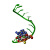
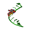
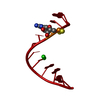
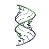
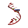
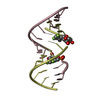
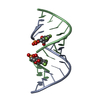
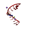
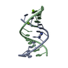
- PDB-2q1r: Crystal Structure Analysis of the RNA Dodecamer CGCGAAUUAGCG, wit... -
+ Open data

- Basic information
| Entry | Database: PDB / ID: 2q1r | ||||||||||||||||||
|---|---|---|---|---|---|---|---|---|---|---|---|---|---|---|---|---|---|---|---|
| Title | Crystal Structure Analysis of the RNA Dodecamer CGCGAAUUAGCG, with a G-A mismatch. | ||||||||||||||||||
 Components Components | RNA (5'-R(* Keywords KeywordsRNA / G-A mismatch / RNAi | Function / homology | RNA / RNA (> 10) |  Function and homology information Function and homology informationMethod |  X-RAY DIFFRACTION / SYNCHROTRON / MOLECULAR REPLACEMENT / Resolution: 1.12 Å X-RAY DIFFRACTION / SYNCHROTRON / MOLECULAR REPLACEMENT / Resolution: 1.12 Å  Authors AuthorsLi, F. / Pallan, P.S. |  CitationJournal: Nucleic Acids Res. / Year: 2007 CitationJournal: Nucleic Acids Res. / Year: 2007Title: Crystal structure, stability and in vitro RNAi activity of oligoribonucleotides containing the ribo-difluorotoluyl nucleotide: insights into substrate requirements by the human RISC Ago2 enzyme. Authors: Li, F. / Pallan, P.S. / Maier, M.A. / Rajeev, K.G. / Mathieu, S.L. / Kreutz, C. / Fan, Y. / Sanghvi, J. / Micura, R. / Rozners, E. / Manoharan, M. / Egli, M. History |
|
- Structure visualization
| Structure viewer | Molecule: MolmilJmol/JSmol |
|---|
- Downloads & links
-Download
| PDBx/mmCIF format | 2q1r.cif.gz | 27.2 KB | Display | PDBx/mmCIF format |
|---|---|---|---|---|
| PDB format | pdb2q1r.ent.gz | 17.9 KB | Display | PDB format |
| PDBx/mmJSON format | 2q1r.json.gz | Tree view | PDBx/mmJSON format | |
| Others |  Other downloads Other downloads |
-Validation report
| Arichive directory | https://data.pdbj.org/pub/pdb/validation_reports/q1/2q1rftp://data.pdbj.org/pub/pdb/validation_reports/q1/2q1r | HTTPS FTP |
|---|
-Related structure data


-Links
 PDBj
PDBj


- Assembly
| Deposited unit | 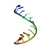
| ||||||||
|---|---|---|---|---|---|---|---|---|---|
| 1 | 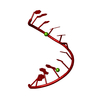
| ||||||||
| Unit cell |
| ||||||||
| Components on special symmetry positions |
| ||||||||
| Details | The biological assembly is a duplex and correspnds to the crystallographic asymmetric unit, hence no symmetry operators are needed |
-Components
| #1: RNA chain | Mass: 3851.360 Da / Num. of mol.: 1 Source method: isolated from a genetically manipulated source Details: Chemically synthesized | ||
|---|---|---|---|
| #2: Chemical |   Mass: 24.305 Da / Num. of mol.: 2 / Source method: obtained synthetically / Formula: Mg Mass: 24.305 Da / Num. of mol.: 2 / Source method: obtained synthetically / Formula: Mg#3: Water | ChemComp-HOH / |  Mass: 18.015 Da / Num. of mol.: 75 / Source method: isolated from a natural source / Formula: H2O Mass: 18.015 Da / Num. of mol.: 75 / Source method: isolated from a natural source / Formula: H2O |
-Experimental details
-Experiment
| Experiment | Method: X-RAY DIFFRACTION / Number of used crystals: 1 |
|---|
- Sample preparation
| Crystal | Density Matthews: 2.33 Å3/Da / Density % sol: 47.12 % | ||||||||||||||||||||||||||||
|---|---|---|---|---|---|---|---|---|---|---|---|---|---|---|---|---|---|---|---|---|---|---|---|---|---|---|---|---|---|
| Crystal grow | Temperature: 291 K / Method: vapor diffusion, hanging drop / pH: 7 Details: Droplets containing 0.5 mM oligonucleotide, 5% MPD, 20 mM sodium cacodylate, pH 7.0, 6 mM spermine-4HCl, 40 mM sodium chloride and 10 mM magnesium chloride were equilibrated against a ...Details: Droplets containing 0.5 mM oligonucleotide, 5% MPD, 20 mM sodium cacodylate, pH 7.0, 6 mM spermine-4HCl, 40 mM sodium chloride and 10 mM magnesium chloride were equilibrated against a reservoir of 35% MPD., VAPOR DIFFUSION, HANGING DROP, temperature 291K | ||||||||||||||||||||||||||||
| Components of the solutions |
|
-Data collection
| Diffraction | Mean temperature: 120 K |
|---|---|
| Diffraction source | Source: SYNCHROTRON / Site: APS  / Beamline: 5ID-B / Wavelength: 0.979 Å / Beamline: 5ID-B / Wavelength: 0.979 Å |
| Detector | Type: MARMOSAIC 225 mm CCD / Detector: CCD / Date: Mar 26, 2004 |
| Radiation | Protocol: SINGLE WAVELENGTH / Monochromatic (M) / Laue (L): M / Scattering type: x-ray |
| Radiation wavelength | Wavelength: 0.979 Å / Relative weight: 1 |
| Reflection | Resolution: 1.12→24.84 Å / Num. all: 13446 / Num. obs: 13280 / % possible obs: 96.4 % / Observed criterion σ(F): 0 / Observed criterion σ(I): 0 / Rmerge(I) obs: 0.035 |
| Reflection shell | Resolution: 1.12→1.16 Å / Rmerge(I) obs: 0.277 / Num. unique all: 1330 / % possible all: 89.3 |
- Processing
| Software |
| |||||||||||||||||||||||||
|---|---|---|---|---|---|---|---|---|---|---|---|---|---|---|---|---|---|---|---|---|---|---|---|---|---|---|
| Refinement | Method to determine structure: MOLECULAR REPLACEMENT Starting model: A native A-RNA Resolution: 1.12→10 Å / Cross valid method: THROUGHOUT / σ(F): 4 / Stereochemistry target values: Engh & Huber
| |||||||||||||||||||||||||
| Refinement step | Cycle: LAST / Resolution: 1.12→10 Å
| |||||||||||||||||||||||||
| Refine LS restraints |
|
