 Movie
Movie Controller

[English] 日本語
 Yorodumi
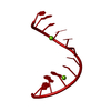
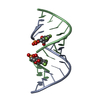
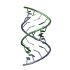
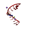
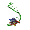
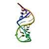
- PDB-4rby: First X-ray structure of RNA containing guanosine phosphorodithioate -
+ Open data

- Basic information
| Entry | Database: PDB / ID: 4rby | ||||||||||||||||||||
|---|---|---|---|---|---|---|---|---|---|---|---|---|---|---|---|---|---|---|---|---|---|
| Title | First X-ray structure of RNA containing guanosine phosphorodithioate | ||||||||||||||||||||
 Components Components | 5'-R(* Keywords KeywordsRNA / phosphorodithioate RNA / guanosine phosphorodithioate / phosphorothioate / guanosine analogue / hydrophobic / backbone | Function / homology | STRONTIUM ION / RNA / RNA (> 10) |  Function and homology information Function and homology informationBiological species | synthetic construct (others) | Method |  X-RAY DIFFRACTION / SYNCHROTRON / MOLECULAR REPLACEMENT / Resolution: 1.19 Å X-RAY DIFFRACTION / SYNCHROTRON / MOLECULAR REPLACEMENT / Resolution: 1.19 Å  Authors AuthorsPallan, P.S. / Egli, M. |  CitationJournal: To be Published CitationJournal: To be PublishedTitle: Crystal Structure, Stability and siRNA Activity of Phosphorodithioate-Modified RNAs Authors: Pallan, P.S. / Yang, X. / Sierant, M. / Abeydeera, N.D. / Hassell, T. / Martinez, C. / Janicka, M. / Nawrot, B. / Egli, M. History |
|
- Structure visualization
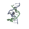
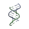
| Structure viewer | Molecule: MolmilJmol/JSmol |
|---|
- Downloads & links
-Download
| PDBx/mmCIF format | 4rby.cif.gz | 20.3 KB | Display | PDBx/mmCIF format |
|---|---|---|---|---|
| PDB format | pdb4rby.ent.gz | 11.6 KB | Display | PDB format |
| PDBx/mmJSON format | 4rby.json.gz | Tree view | PDBx/mmJSON format | |
| Others |  Other downloads Other downloads |
-Validation report
| Arichive directory | https://data.pdbj.org/pub/pdb/validation_reports/rb/4rbyftp://data.pdbj.org/pub/pdb/validation_reports/rb/4rby | HTTPS FTP |
|---|
-Related structure data
| Related structure data | 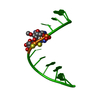 4rbzC  4rc0C  2q1rS C: citing same article ( S: Starting model for refinement |
|---|---|
| Similar structure data |

-Links
 PDBj
PDBj


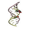
- Assembly
| Deposited unit | 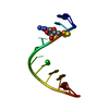
| |||||||||||||||
|---|---|---|---|---|---|---|---|---|---|---|---|---|---|---|---|---|
| 1 | 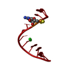
| |||||||||||||||
| Unit cell |
| |||||||||||||||
| Components on special symmetry positions |
|
-Components
| #1: RNA chain | Mass: 3885.507 Da / Num. of mol.: 1 / Source method: obtained synthetically / Details: Guanosine phosphorodithioate RNA / Source: (synth.) synthetic construct (others) | ||
|---|---|---|---|
| #2: Chemical |   Mass: 87.620 Da / Num. of mol.: 2 / Source method: obtained synthetically / Formula: Sr Mass: 87.620 Da / Num. of mol.: 2 / Source method: obtained synthetically / Formula: Sr#3: Water | ChemComp-HOH / |  Mass: 18.015 Da / Num. of mol.: 100 / Source method: isolated from a natural source / Formula: H2O Mass: 18.015 Da / Num. of mol.: 100 / Source method: isolated from a natural source / Formula: H2O |
-Experimental details
-Experiment
| Experiment | Method: X-RAY DIFFRACTION / Number of used crystals: 1 |
|---|
- Sample preparation
| Crystal | Density Matthews: 2.29 Å3/Da / Density % sol: 46.3 % |
|---|---|
| Crystal grow | Temperature: 291 K / Method: vapor diffusion, hanging drop / pH: 7 Details: 20 mM sodium cacodylate, 40 mM sodium chloride, 10 mM barium chloride, 6 mM spermine tetrahydrochloride, 5% v/v MPD, pH 7.0, VAPOR DIFFUSION, HANGING DROP, temperature 291K |
-Data collection
| Diffraction | Mean temperature: 100 K |
|---|---|
| Diffraction source | Source: SYNCHROTRON / Site: APS  / Beamline: 21-ID-F / Wavelength: 0.97872 Å / Beamline: 21-ID-F / Wavelength: 0.97872 Å |
| Detector | Type: MARMOSAIC 325 mm CCD / Detector: CCD / Date: Oct 11, 2012 |
| Radiation | Monochromator: diamond(111) / Protocol: SINGLE WAVELENGTH / Monochromatic (M) / Laue (L): M / Scattering type: x-ray |
| Radiation wavelength | Wavelength: 0.97872 Å / Relative weight: 1 |
| Reflection | Resolution: 1.19→50 Å / Num. all: 11477 / Num. obs: 11213 / % possible obs: 97.7 % / Observed criterion σ(I): 5 / Redundancy: 7 % / Rmerge(I) obs: 0.044 / Net I/σ(I): 16.49 |
| Reflection shell | Resolution: 1.19→1.23 Å / Redundancy: 5.2 % / Rmerge(I) obs: 0.037 / Mean I/σ(I) obs: 20.71 / Num. unique all: 1012 / % possible all: 88.5 |
- Processing
| Software |
| ||||||||||||||||||||
|---|---|---|---|---|---|---|---|---|---|---|---|---|---|---|---|---|---|---|---|---|---|
| Refinement | Method to determine structure: MOLECULAR REPLACEMENT Starting model: PDB ENTRY 2Q1R Resolution: 1.19→50 Å / Stereochemistry target values: Engh & Huber
| ||||||||||||||||||||
| Refinement step | Cycle: LAST / Resolution: 1.19→50 Å
| ||||||||||||||||||||
| Refine LS restraints |
|
