 Movie
Movie Controller

[English] 日本語
 Yorodumi
- PDB-2q00: Crystal structure of the P95883_SULSO protein from Sulfolobus sol... -
+ Open data

- Basic information
| Entry | Database: PDB / ID: 2q00 | ||||||
|---|---|---|---|---|---|---|---|
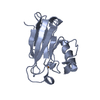
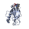
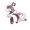
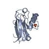
| Title | Crystal structure of the P95883_SULSO protein from Sulfolobus solfataricus. NESG target SsR10. | ||||||
 Components Components | Orf c02003 protein | ||||||
 Keywords Keywords | STRUCTURAL GENOMICS / UNKNOWN FUNCTION / P95883 / NESG / SSO2109 / PSI-2 / Protein Structure Initiative / Northeast Structural Genomics Consortium | ||||||
| Function / homology | Archaeal PaREP1 / Archaeal PaREP1/PaREP8 family / Nucleotidyltransferases domain 2 / Four Helix Bundle (Hemerythrin (Met), subunit A) / Up-down Bundle / Mainly Alpha / Orf c02003 protein Function and homology information Function and homology information | ||||||
| Biological species |   Sulfolobus solfataricus P2 (archaea) Sulfolobus solfataricus P2 (archaea) | ||||||
| Method |  X-RAY DIFFRACTION / SYNCHROTRON / SIRAS / Resolution: 2.4 Å X-RAY DIFFRACTION / SYNCHROTRON / SIRAS / Resolution: 2.4 Å | ||||||
 Authors Authors | Vorobiev, S.M. / Chen, Y. / Seetharaman, J. / Wang, D. / Owens, L. / Ma, L.-C. / Cunningham, K. / Fang, Y. / Xiao, R. / Acton, T.B. ...Vorobiev, S.M. / Chen, Y. / Seetharaman, J. / Wang, D. / Owens, L. / Ma, L.-C. / Cunningham, K. / Fang, Y. / Xiao, R. / Acton, T.B. / Montelione, G.T. / Tong, L. / Hunt, J.F. / Northeast Structural Genomics Consortium (NESG) | ||||||
 Citation Citation | Journal: To be Published Title: Crystal structure of the P95883_SULSO protein from Sulfolobus solfataricus. Authors: Vorobiev, S.M. / Chen, Y. / Seetharaman, J. / Wang, D. / Owens, L. / Ma, L.-C. / Cunningham, K. / Fang, Y. / Xiao, R. / Acton, T.B. / Montelione, G.T. / Tong, L. / Hunt, J.F. | ||||||
| History |
|
- Structure visualization
| Structure viewer | Molecule: MolmilJmol/JSmol |
|---|
- Downloads & links
-Download
| PDBx/mmCIF format | 2q00.cif.gz | 58.6 KB | Display | PDBx/mmCIF format |
|---|---|---|---|---|
| PDB format | pdb2q00.ent.gz | 43.9 KB | Display | PDB format |
| PDBx/mmJSON format | 2q00.json.gz | Tree view | PDBx/mmJSON format | |
| Others |  Other downloads Other downloads |
-Validation report
| Arichive directory | https://data.pdbj.org/pub/pdb/validation_reports/q0/2q00ftp://data.pdbj.org/pub/pdb/validation_reports/q0/2q00 | HTTPS FTP |
|---|
-Related structure data
| Similar structure data | |
|---|---|
| Other databases |


-Links
 PDBj
PDBj- Assembly
| Deposited unit | 
| ||||||||
|---|---|---|---|---|---|---|---|---|---|
| 1 | 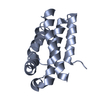
| ||||||||
| 2 | 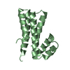
| ||||||||
| Unit cell |
|
-Components
| #1: Protein | Mass: 15084.201 Da / Num. of mol.: 2 Source method: isolated from a genetically manipulated source Source: (gene. exp.) Sulfolobus solfataricus P2 (archaea) / Species: Sulfolobus solfataricus / Strain: P2, DSM 1617, JCM 11322 / Gene: orf c02003, SSO2109 / Plasmid: pET21 / Production host:  #2: Water | ChemComp-HOH / |  Mass: 18.015 Da / Num. of mol.: 29 / Source method: isolated from a natural source / Formula: H2O Mass: 18.015 Da / Num. of mol.: 29 / Source method: isolated from a natural source / Formula: H2O |
|---|
-Experimental details
-Experiment
| Experiment | Method: X-RAY DIFFRACTION / Number of used crystals: 1 |
|---|
- Sample preparation
| Crystal | Density Matthews: 2.83 Å3/Da / Density % sol: 56.58 % Description: THE STRUCTURE FACTOR FILE CONTAINS FRIEDEL PAIRS |
|---|---|
| Crystal grow | Temperature: 277 K / Method: microbatch under oil / pH: 6 Details: 20 % PEG 4000, 0.1 M Ammonium thiocyanate, 0.1 M MES, pH 6.0, MICROBATCH UNDER OIL, temperature 277K |
-Data collection
| Diffraction | Mean temperature: 100 K |
|---|---|
| Diffraction source | Source: SYNCHROTRON / Site: NSLS  / Beamline: X4A / Wavelength: 0.97909 Å / Beamline: X4A / Wavelength: 0.97909 Å |
| Detector | Type: ADSC QUANTUM 4 / Detector: CCD / Date: May 1, 2007 |
| Radiation | Protocol: SINGLE WAVELENGTH / Monochromatic (M) / Laue (L): M / Scattering type: x-ray |
| Radiation wavelength | Wavelength: 0.97909 Å / Relative weight: 1 |
| Reflection | Resolution: 2.4→50 Å / Num. all: 25384 / Num. obs: 25384 / % possible obs: 97.4 % / Observed criterion σ(F): 0 / Observed criterion σ(I): 0 / Redundancy: 3.8 % / Biso Wilson estimate: 20.4 Å2 / Rmerge(I) obs: 0.053 / Net I/σ(I): 26.8 |
| Reflection shell | Resolution: 2.4→2.5 Å / Redundancy: 3.8 % / Rmerge(I) obs: 0.545 / Mean I/σ(I) obs: 2.6 / Num. unique all: 5179 / % possible all: 100 |
- Processing
| Software |
| |||||||||||||||||||||
|---|---|---|---|---|---|---|---|---|---|---|---|---|---|---|---|---|---|---|---|---|---|---|
| Refinement | Method to determine structure: SIRAS / Resolution: 2.4→36.03 Å / Rfactor Rfree error: 0.009 / Data cutoff high absF: 418136.47 / Data cutoff low absF: 0 / Isotropic thermal model: RESTRAINED / Cross valid method: THROUGHOUT / σ(F): 2 / Stereochemistry target values: Engh & Huber / Details: THE FRIEDEL PAIRS WERE USED FOR PHASING
| |||||||||||||||||||||
| Solvent computation | Solvent model: FLAT MODEL / Bsol: 42.509 Å2 / ksol: 0.30844 e/Å3 | |||||||||||||||||||||
| Displacement parameters | Biso mean: 57.3 Å2
| |||||||||||||||||||||
| Refine analyze |
| |||||||||||||||||||||
| Refinement step | Cycle: LAST / Resolution: 2.4→36.03 Å
| |||||||||||||||||||||
| Refine LS restraints |
| |||||||||||||||||||||
| LS refinement shell | Resolution: 2.4→2.55 Å / Rfactor Rfree error: 0.023 / Total num. of bins used: 6
| |||||||||||||||||||||
| Xplor file |
|
