 Movie
Movie Controller


+ Open data

- Basic information
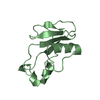
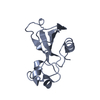
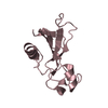
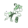
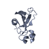
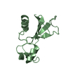
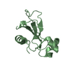
| Entry | Database: PDB / ID: 2f56 | ||||||
|---|---|---|---|---|---|---|---|
| Title | Barnase cross-linked with glutaraldehyde soaked in 6M urea | ||||||
 Components Components | Ribonuclease | ||||||
 Keywords Keywords | HYDROLASE / DENATURATION / LYSOZYME / BARNASE / CROSS-LINKED CRYSTALS / GLUTARALDEHYDE / UREA / THIOUREA / BROMOETHANOL | ||||||
| Function / homology |  Function and homology information Function and homology informationHydrolases; Acting on ester bonds; Endoribonucleases producing 3'-phosphomonoesters / RNA endonuclease activity / hydrolase activity / RNA binding / extracellular region Similarity search - Function | ||||||
| Biological species |  | ||||||
| Method |  X-RAY DIFFRACTION / SYNCHROTRON / FOURIER SYNTHESIS / Resolution: 1.955 Å X-RAY DIFFRACTION / SYNCHROTRON / FOURIER SYNTHESIS / Resolution: 1.955 Å | ||||||
 Authors Authors | Prange, T. / Salem, M. | ||||||
 Citation Citation | Journal: Biochim.Biophys.Acta / Year: 2006 Title: On the edge of the denaturation process: Application of X-ray diffraction to barnase and lysozyme cross-linked crystals with denaturants in molar concentrations. Authors: Salem, M. / Mauguen, Y. / Prange, T. | ||||||
| History |
|
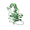
- Structure visualization
| Structure viewer | Molecule: MolmilJmol/JSmol |
|---|
- Downloads & links
-Download
| PDBx/mmCIF format | 2f56.cif.gz | 83.7 KB | Display | PDBx/mmCIF format |
|---|---|---|---|---|
| PDB format | pdb2f56.ent.gz | 62 KB | Display | PDB format |
| PDBx/mmJSON format | 2f56.json.gz | Tree view | PDBx/mmJSON format | |
| Others |  Other downloads Other downloads |
-Validation report
| Arichive directory | https://data.pdbj.org/pub/pdb/validation_reports/f5/2f56ftp://data.pdbj.org/pub/pdb/validation_reports/f5/2f56 | HTTPS FTP |
|---|
-Related structure data
| Related structure data |  2f2nC  2f30C 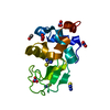 2f4aC 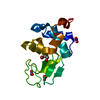 2f4gC  2f4ySC  2f5mC  2f5wC S: Starting model for refinement C: citing same article ( |
|---|---|
| Similar structure data |


-Links
 PDBj
PDBj
- Assembly
| Deposited unit | 
| ||||||||
|---|---|---|---|---|---|---|---|---|---|
| 1 | 
| ||||||||
| 2 | 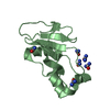
| ||||||||
| 3 | 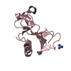
| ||||||||
| 4 | 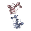
| ||||||||
| Unit cell |
|
-Components
| #1: Protein | Mass: 12199.515 Da / Num. of mol.: 3 Source method: isolated from a genetically manipulated source Source: (gene. exp.) References: UniProt: P00648, Hydrolases; Acting on ester bonds; Endoribonucleases producing 3'-phosphomonoesters #2: Chemical | ChemComp-URE /   Mass: 60.055 Da / Num. of mol.: 11 / Source method: obtained synthetically / Formula: CH4N2O Mass: 60.055 Da / Num. of mol.: 11 / Source method: obtained synthetically / Formula: CH4N2O#3: Chemical | ChemComp-ZN / |   Mass: 65.409 Da / Num. of mol.: 1 / Source method: obtained synthetically / Formula: Zn Mass: 65.409 Da / Num. of mol.: 1 / Source method: obtained synthetically / Formula: Zn#4: Water | ChemComp-HOH / |  Mass: 18.015 Da / Num. of mol.: 236 / Source method: isolated from a natural source / Formula: H2O Mass: 18.015 Da / Num. of mol.: 236 / Source method: isolated from a natural source / Formula: H2O |
|---|
-Experimental details
-Experiment
| Experiment | Method: X-RAY DIFFRACTION / Number of used crystals: 1 |
|---|
- Sample preparation
| Crystal | Density Matthews: 2.26 Å3/Da / Density % sol: 45.57 % |
|---|---|
| Crystal grow | Temperature: 300 K / Method: vapor diffusion, hanging drop / pH: 8 Details: AMMONIUM SULFATE, HEPES BUFFER. PROTEIN 20 MG/ML, pH 8, VAPOR DIFFUSION, HANGING DROP, temperature 300K |
-Data collection
| Diffraction | Mean temperature: 277 K |
|---|---|
| Diffraction source | Source: SYNCHROTRON / Site: LURE  / Beamline: DW32 / Wavelength: 0.964 Å / Beamline: DW32 / Wavelength: 0.964 Å |
| Detector | Type: MARRESEARCH / Detector: IMAGE PLATE / Date: Mar 25, 2002 / Details: CURVATED MULTILAYER MIRROR |
| Radiation | Monochromator: CURVATED SI(111) / Protocol: SINGLE WAVELENGTH / Monochromatic (M) / Laue (L): M / Scattering type: x-ray |
| Radiation wavelength | Wavelength: 0.964 Å / Relative weight: 1 |
| Reflection | Resolution: 1.955→10 Å / Num. all: 23026 / Num. obs: 21767 / % possible obs: 99.2 % / Observed criterion σ(F): 2 / Observed criterion σ(I): 4 / Net I/σ(I): 22 |
| Reflection shell | Resolution: 1.955→2.1 Å / Mean I/σ(I) obs: 10.7 / % possible all: 99.9 |
- Processing
| Software |
| ||||||||||||||||||||||||||||||||||||
|---|---|---|---|---|---|---|---|---|---|---|---|---|---|---|---|---|---|---|---|---|---|---|---|---|---|---|---|---|---|---|---|---|---|---|---|---|---|
| Refinement | Method to determine structure: FOURIER SYNTHESIS Starting model: PDB ENTRY 2F4Y Resolution: 1.955→10 Å / Num. parameters: 11467 / Num. restraintsaints: 10750 / σ(F): 4 / Stereochemistry target values: ENGH AND HUBER
| ||||||||||||||||||||||||||||||||||||
| Refine analyze | Num. disordered residues: 0 / Occupancy sum hydrogen: 2442 / Occupancy sum non hydrogen: 2840 | ||||||||||||||||||||||||||||||||||||
| Refinement step | Cycle: LAST / Resolution: 1.955→10 Å
| ||||||||||||||||||||||||||||||||||||
| Refine LS restraints |
| ||||||||||||||||||||||||||||||||||||
| LS refinement shell |
|
