 Movie
Movie Controller


+ Open data

- Basic information
| Entry | Database: PDB / ID: 2pzx | ||||||
|---|---|---|---|---|---|---|---|
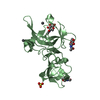
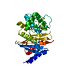
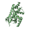
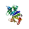
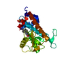
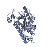
| Title | Structure of the methuselah ectodomain with peptide inhibitor | ||||||
 Components Components | G-protein coupled receptor Mth | ||||||
 Keywords Keywords | SIGNALING PROTEIN / GPCR G protein-coupled receptor ectodomain | ||||||
| Function / homology |  Function and homology information Function and homology informationresponse to paraquat / G protein-coupled peptide receptor activity / response to starvation / synaptic vesicle exocytosis / peptide binding / response to reactive oxygen species / determination of adult lifespan / G protein-coupled receptor activity / presynapse / response to heat ...response to paraquat / G protein-coupled peptide receptor activity / response to starvation / synaptic vesicle exocytosis / peptide binding / response to reactive oxygen species / determination of adult lifespan / G protein-coupled receptor activity / presynapse / response to heat / cell surface receptor signaling pathway / G protein-coupled receptor signaling pathway / membrane / plasma membrane Similarity search - Function | ||||||
| Biological species |  | ||||||
| Method |  X-RAY DIFFRACTION / SYNCHROTRON / MOLECULAR REPLACEMENT / Resolution: 3.5 Å X-RAY DIFFRACTION / SYNCHROTRON / MOLECULAR REPLACEMENT / Resolution: 3.5 Å | ||||||
 Authors Authors | West Jr., A.P. / Ja, W.W. / Delker, S.L. / Bjorkman, P.J. / Benzer, S. / Roberts, R.W. | ||||||
 Citation Citation | Journal: Nat.Chem.Biol. / Year: 2007 Title: Extension of Drosophila melanogaster life span with a GPCR peptide inhibitor. Authors: Ja, W.W. / West, A.P. / Delker, S.L. / Bjorkman, P.J. / Benzer, S. / Roberts, R.W. | ||||||
| History |
|
- Structure visualization
| Structure viewer | Molecule: MolmilJmol/JSmol |
|---|
- Downloads & links
-Download
| PDBx/mmCIF format | 2pzx.cif.gz | 211.4 KB | Display | PDBx/mmCIF format |
|---|---|---|---|---|
| PDB format | pdb2pzx.ent.gz | 178.6 KB | Display | PDB format |
| PDBx/mmJSON format | 2pzx.json.gz | Tree view | PDBx/mmJSON format | |
| Others |  Other downloads Other downloads |
-Validation report
| Summary document | 2pzx_validation.pdf.gz | 455.7 KB | Display | wwPDB validaton report |
|---|---|---|---|---|
| Full document | 2pzx_full_validation.pdf.gz | 525.8 KB | Display | |
| Data in XML | 2pzx_validation.xml.gz | 40.5 KB | Display | |
| Data in CIF | 2pzx_validation.cif.gz | 54 KB | Display | |
| Arichive directory | https://data.pdbj.org/pub/pdb/validation_reports/pz/2pzxftp://data.pdbj.org/pub/pdb/validation_reports/pz/2pzx | HTTPS FTP |
-Related structure data
| Related structure data |  1fjrS S: Starting model for refinement |
|---|---|
| Similar structure data |


-Links
 PDBj
PDBj
- Assembly
| Deposited unit | 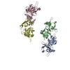
| ||||||||
|---|---|---|---|---|---|---|---|---|---|
| 1 | 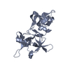
| ||||||||
| 2 | 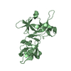
| ||||||||
| 3 | 
| ||||||||
| 4 | 
| ||||||||
| Unit cell |
|
-Components
| #1: Protein | Mass: 21835.809 Da / Num. of mol.: 4 / Fragment: N-terminal extracellular domain Source method: isolated from a genetically manipulated source Source: (gene. exp.)  unidentified baculovirus / References: UniProt: O97148 unidentified baculovirus / References: UniProt: O97148Has protein modification | Y | |
|---|
-Experimental details
-Experiment
| Experiment | Method: X-RAY DIFFRACTION / Number of used crystals: 1 |
|---|
- Sample preparation
| Crystal | Density Matthews: 4.42 Å3/Da / Density % sol: 72.15 % |
|---|---|
| Crystal grow | Temperature: 298 K / pH: 7.5 Details: 1.7 M ammonium sulfate 2% w/v PEG 400 0.1 M HEPES-KOH pH 7.5, VAPOR DIFFUSION, HANGING DROP, temperature 298K, pH 7.50 |
-Data collection
| Diffraction | Mean temperature: 100 K | ||||||||||||||||||||||||||||||||||||||||||||||||||||||||||||||||||
|---|---|---|---|---|---|---|---|---|---|---|---|---|---|---|---|---|---|---|---|---|---|---|---|---|---|---|---|---|---|---|---|---|---|---|---|---|---|---|---|---|---|---|---|---|---|---|---|---|---|---|---|---|---|---|---|---|---|---|---|---|---|---|---|---|---|---|---|
| Diffraction source | Source: SYNCHROTRON / Site: ALS  / Beamline: 8.2.2 / Wavelength: 1.0781 / Beamline: 8.2.2 / Wavelength: 1.0781 | ||||||||||||||||||||||||||||||||||||||||||||||||||||||||||||||||||
| Detector | Type: ADSC QUANTUM 4 / Detector: CCD / Date: Apr 4, 2004 | ||||||||||||||||||||||||||||||||||||||||||||||||||||||||||||||||||
| Radiation | Protocol: SINGLE WAVELENGTH / Monochromatic (M) / Laue (L): M / Scattering type: x-ray | ||||||||||||||||||||||||||||||||||||||||||||||||||||||||||||||||||
| Radiation wavelength | Wavelength: 1.0781 Å / Relative weight: 1 | ||||||||||||||||||||||||||||||||||||||||||||||||||||||||||||||||||
| Reflection | Av σ(I) over netI: 6.2 / Number: 66512 / Rmerge(I) obs: 0.155 / Χ2: 1.33 / D res high: 3.5 Å / D res low: 20 Å / Num. obs: 18125 / % possible obs: 95.3 | ||||||||||||||||||||||||||||||||||||||||||||||||||||||||||||||||||
| Diffraction reflection shell |
| ||||||||||||||||||||||||||||||||||||||||||||||||||||||||||||||||||
| Reflection | Resolution: 3.5→20 Å / Num. obs: 18125 / % possible obs: 95.3 % / Rmerge(I) obs: 0.155 / Net I/σ(I): 6.2 | ||||||||||||||||||||||||||||||||||||||||||||||||||||||||||||||||||
| Reflection shell | Resolution: 3.5→3.62 Å / Rmerge(I) obs: 0.37 / % possible all: 96.9 |
- Processing
| Software |
| ||||||||||||||||||||||||||||||||||||||||||||||||||||||||||||
|---|---|---|---|---|---|---|---|---|---|---|---|---|---|---|---|---|---|---|---|---|---|---|---|---|---|---|---|---|---|---|---|---|---|---|---|---|---|---|---|---|---|---|---|---|---|---|---|---|---|---|---|---|---|---|---|---|---|---|---|---|---|
| Refinement | Method to determine structure: MOLECULAR REPLACEMENT Starting model: PDB ENTRY 1FJR Resolution: 3.5→7.99 Å / Rfactor Rfree error: 0.014 / Data cutoff high absF: 1208292.02 / Data cutoff low absF: 0 / Isotropic thermal model: RESTRAINED / Cross valid method: THROUGHOUT / σ(F): 0 / Stereochemistry target values: ENGH & HUBER Details: THE R8-01 15-MER PEPTIDE (FPSSWLQRYYLAKRR) WAS NOT INCLUDED IN THE MODEL. THIS CRYSTAL HAS A STACKING FAULT-TYPE DISORDER, WHICH CAN ONLY BE PARTIALLY MODELED. THE OVERLAPPING NCS-RELATED ...Details: THE R8-01 15-MER PEPTIDE (FPSSWLQRYYLAKRR) WAS NOT INCLUDED IN THE MODEL. THIS CRYSTAL HAS A STACKING FAULT-TYPE DISORDER, WHICH CAN ONLY BE PARTIALLY MODELED. THE OVERLAPPING NCS-RELATED MOLECULES ARE NECESSARY TO MODEL THE DISORDER. Refinement was done with 8 strict NCS matrices as follows: matrix_1 = ( 1.000000 0.000000 0.000000 ) ( 0.000000 1.000000 0.000000 ) ( 0.000000 0.000000 1.000000 ) vector_1 = ( 0.0000 0.0000 0.0000 ) matrix_2 = ( -0.025450 -0.999672 -0.002776 ) ( -0.999629 0.025476 -0.009636 ) ( 0.009703 0.002530 -0.999950 ) vector_2 = ( 95.7336 93.6049 30.5814 ) matrix_3 = ( 0.999874 -0.015577 -0.003041 ) ( -0.015587 -0.999874 -0.003124 ) ( -0.002992 0.003171 -0.999991 ) vector_3 = ( -46.4752 48.4846 0.7945 ) matrix_4 = ( -0.030796 -0.999466 -0.010894 ) ( 0.999470 -0.030907 0.010218 ) ( -0.010550 -0.010574 0.999888 ) vector_4 = ( 48.9629 -45.9887 -29.5468 ) matrix_5 = ( -0.010011 -0.999911 -0.008770 ) ( 0.999838 -0.010141 0.014863 ) ( -0.014951 -0.008620 0.999851 ) vector_5 = ( 48.0912 -47.3711 31.8114 ) matrix_6 = ( 0.999542 -0.030236 -0.001427 ) ( -0.030235 -0.999542 0.001202 ) ( -0.001463 -0.001158 -0.999998 ) vector_6 = ( -45.5886 48.9605 61.9827 ) matrix_7 = ( 1.000000 0.000000 0.000000 ) ( 0.000000 1.000000 0.000000 ) ( 0.000000 0.000000 1.000000 ) vector_7 = ( 0.0000 0.0000 0.0000 ) matrix_8 = ( -0.025450 -0.999672 -0.002776 ) ( -0.999629 0.025476 -0.009636 ) ( 0.009703 0.002530 -0.999950 ) vector_8 = ( 95.7336 93.6049 30.5814 ). Author has stated one complication: Because of the stacking fault-type disorder, the alternate conformers in the current PDB file were assigned different chain IDs during author's refinement. The two alternate conformers of chain C were assigned chain IDs C and F, and the two conformers of chain D were assigned D and E. During the processing of deposition, alternate conformers were used because of the essential PDB format requirement. Therefore, during author's refinement, there were six chains A/B/C/D/E/F. To get the right occupancies during the refinement (1.0 on chains A/B, 0.5 on chains C/D/E/F), author set the occupancies of the starting file at 0.5, and put in two copies of the ncs matrices for the chains A and B with occupancy of 1.0. So there are 8 matrices.
| ||||||||||||||||||||||||||||||||||||||||||||||||||||||||||||
| Solvent computation | Solvent model: FLAT MODEL / Bsol: 21.5771 Å2 / ksol: 0.172883 e/Å3 | ||||||||||||||||||||||||||||||||||||||||||||||||||||||||||||
| Displacement parameters | Biso mean: 53.3 Å2
| ||||||||||||||||||||||||||||||||||||||||||||||||||||||||||||
| Refine analyze |
| ||||||||||||||||||||||||||||||||||||||||||||||||||||||||||||
| Refinement step | Cycle: LAST / Resolution: 3.5→7.99 Å
| ||||||||||||||||||||||||||||||||||||||||||||||||||||||||||||
| Refine LS restraints |
| ||||||||||||||||||||||||||||||||||||||||||||||||||||||||||||
| Refine LS restraints NCS | NCS model details: CONSTR | ||||||||||||||||||||||||||||||||||||||||||||||||||||||||||||
| LS refinement shell | Resolution: 3.5→3.7 Å / Rfactor Rfree error: 0.037 / Total num. of bins used: 6
| ||||||||||||||||||||||||||||||||||||||||||||||||||||||||||||
| Xplor file | Serial no: 1 / Param file: protein_rep.param / Topol file: protein.top |
