Method to determine structure: MOLECULAR REPLACEMENT / Resolution: 1.5→53.15 Å / Cor.coef. Fo:Fc: 0.969 / Cor.coef. Fo:Fc free: 0.944 / SU B: 3.664 / SU ML: 0.061 / Cross valid method: THROUGHOUT / σ(F): 0 / σ(I): 2 / ESU R: 0.102 / ESU R Free: 0.088 / Stereochemistry target values: MAXIMUM LIKELIHOOD Details: HYDROGENS HAVE BEEN ADDED IN THE RIDING POSITIONS. Authors also state that The structure 2PXR was determined by X-ray crystallography from crystals of CA146 that were grown in the presence ...Details: HYDROGENS HAVE BEEN ADDED IN THE RIDING POSITIONS. Authors also state that The structure 2PXR was determined by X-ray crystallography from crystals of CA146 that were grown in the presence of CAP-1. CAP-1 is not visible in electron density maps, although NMR data provided ligand-protein NOEs that allowed them to build a joint refined structure 2JPR.
Rfactor
Num. reflection
% reflection
Selection details
Rfree
0.221
1107
5.1 %
RANDOM
Rwork
0.163
-
-
-
all
0.166
22915
-
-
obs
0.166
21916
95.2 %
-
Solvent computation
Ion probe radii: 0.8 Å / Shrinkage radii: 0.8 Å / VDW probe radii: 1.4 Å / Solvent model: MASK
Displacement parameters
Biso mean: 24.635 Å2
Baniso -1
Baniso -2
Baniso -3
1-
0.9 Å2
0 Å2
0 Å2
2-
-
-1.16 Å2
0 Å2
3-
-
-
0.26 Å2
Refinement step
Cycle: LAST / Resolution: 1.5→53.15 Å
Protein
Nucleic acid
Ligand
Solvent
Total
Num. atoms
1129
0
3
179
1311
Refine LS restraints
Refine-ID
Type
Dev ideal
Dev ideal target
Number
X-RAY DIFFRACTION
r_bond_refined_d
0.02
0.021
1223
X-RAY DIFFRACTION
r_angle_refined_deg
1.873
1.927
1666
X-RAY DIFFRACTION
r_dihedral_angle_1_deg
5.519
5
148
X-RAY DIFFRACTION
r_dihedral_angle_2_deg
37.792
25.088
57
X-RAY DIFFRACTION
r_dihedral_angle_3_deg
14.835
15
224
X-RAY DIFFRACTION
r_dihedral_angle_4_deg
20.499
15
7
X-RAY DIFFRACTION
r_chiral_restr
0.13
0.2
185
X-RAY DIFFRACTION
r_gen_planes_refined
0.009
0.02
925
X-RAY DIFFRACTION
r_nbd_refined
0.239
0.2
618
X-RAY DIFFRACTION
r_nbtor_refined
0.31
0.2
864
X-RAY DIFFRACTION
r_xyhbond_nbd_refined
0.188
0.2
127
X-RAY DIFFRACTION
r_symmetry_vdw_refined
0.262
0.2
68
X-RAY DIFFRACTION
r_symmetry_hbond_refined
0.239
0.2
23
X-RAY DIFFRACTION
r_symmetry_metal_ion_refined
0.063
0.2
3
X-RAY DIFFRACTION
r_mcbond_it
2.024
1.5
764
X-RAY DIFFRACTION
r_mcangle_it
2.961
2
1225
X-RAY DIFFRACTION
r_scbond_it
3.757
3
520
X-RAY DIFFRACTION
r_scangle_it
5.544
4.5
441
X-RAY DIFFRACTION
r_rigid_bond_restr
2.506
3
1284
X-RAY DIFFRACTION
r_sphericity_free
8.637
3
182
X-RAY DIFFRACTION
r_sphericity_bonded
7.007
3
1192
LS refinement shell
Resolution: 1.5→1.539 Å / Total num. of bins used: 20
Rfactor
Num. reflection
% reflection
Rfree
0.372
63
-
Rwork
0.247
1146
-
obs
-
1209
72.48 %
+
About Yorodumi
-
News
-
Feb 9, 2022. New format data for meta-information of EMDB entries
New format data for meta-information of EMDB entries
Version 3 of the EMDB header file is now the official format.
The previous official version 1.9 will be removed from the archive.
In the structure databanks used in Yorodumi, some data are registered as the other names, "COVID-19 virus" and "2019-nCoV". Here are the details of the virus and the list of structure data.
Jan 31, 2019. EMDB accession codes are about to change! (news from PDBe EMDB page)
EMDB accession codes are about to change! (news from PDBe EMDB page)
The allocation of 4 digits for EMDB accession codes will soon come to an end. Whilst these codes will remain in use, new EMDB accession codes will include an additional digit and will expand incrementally as the available range of codes is exhausted. The current 4-digit format prefixed with “EMD-” (i.e. EMD-XXXX) will advance to a 5-digit format (i.e. EMD-XXXXX), and so on. It is currently estimated that the 4-digit codes will be depleted around Spring 2019, at which point the 5-digit format will come into force.
The EM Navigator/Yorodumi systems omit the EMD- prefix.
Related info.:Q: What is EMD? / ID/Accession-code notation in Yorodumi/EM Navigator
Yorodumi is a browser for structure data from EMDB, PDB, SASBDB, etc.
This page is also the successor to EM Navigator detail page, and also detail information page/front-end page for Omokage search.
The word "yorodu" (or yorozu) is an old Japanese word meaning "ten thousand". "mi" (miru) is to see.
Related info.:EMDB / PDB / SASBDB / Comparison of 3 databanks / Yorodumi Search / Aug 31, 2016. New EM Navigator & Yorodumi / Yorodumi Papers / Jmol/JSmol / Function and homology information / Changes in new EM Navigator and Yorodumi
 Movie
Movie Controller


 Open data
 Basic information
 Components
 Keywords
 Function and homology information
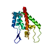
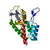
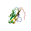
 Human immunodeficiency virus 1
 X-RAY DIFFRACTION /
  Authors
 Citation
 Structure visualization
 Downloads & links
 Other downloads


 PDBj
PDBj

 Assembly


 Mass: 35.453 Da / Num. of mol.: 1 / Source method: obtained synthetically / Formula: Cl
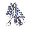
 Mass: 65.409 Da / Num. of mol.: 2 / Source method: obtained synthetically / Formula: Zn
 Mass: 18.015 Da / Num. of mol.: 179 / Source method: isolated from a natural source / Formula: H2O
 Sample preparation
 / Beamline: X12B / Wavelength: 1 Å
 Processing