 Movie
Movie Controller

[English] 日本語
 Yorodumi
- PDB-2pxb: Variant 2 of Ribonucleoprotein Core of the E. Coli Signal Recogni... -
+ Open data

- Basic information
| Entry | Database: PDB / ID: 2pxb | ||||||
|---|---|---|---|---|---|---|---|
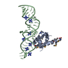
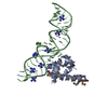
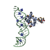
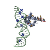
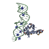
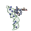
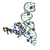
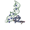
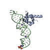
| Title | Variant 2 of Ribonucleoprotein Core of the E. Coli Signal Recognition Particle | ||||||
 Components Components |
| ||||||
 Keywords Keywords | SIGNALING PROTEIN/RNA / GU PAIR / HEXAMINE / RNA PHASING / RNA / CATION BINDING / SIGNALING PROTEIN-RNA COMPLEX | ||||||
| Function / homology |  Function and homology information Function and homology informationsignal recognition particle / signal-recognition-particle GTPase / 7S RNA binding / SRP-dependent cotranslational protein targeting to membrane / protein targeting to membrane / ribonucleoprotein complex / GTPase activity / GTP binding / ATP hydrolysis activity / cytosol Similarity search - Function | ||||||
| Biological species |  | ||||||
| Method |  X-RAY DIFFRACTION / MOLECULAR REPLACEMENT / Resolution: 2.5 Å X-RAY DIFFRACTION / MOLECULAR REPLACEMENT / Resolution: 2.5 Å | ||||||
 Authors Authors | Keel, A.Y. / Rambo, R.P. / Batey, R.T. / Kieft, J.S. | ||||||
 Citation Citation | Journal: Structure / Year: 2007 Title: A General Strategy to Solve the Phase Problem in RNA Crystallography. Authors: Keel, A.Y. / Rambo, R.P. / Batey, R.T. / Kieft, J.S. | ||||||
| History |
|
- Structure visualization
| Structure viewer | Molecule: MolmilJmol/JSmol |
|---|
- Downloads & links
-Download
| PDBx/mmCIF format | 2pxb.cif.gz | 57.5 KB | Display | PDBx/mmCIF format |
|---|---|---|---|---|
| PDB format | pdb2pxb.ent.gz | 39.1 KB | Display | PDB format |
| PDBx/mmJSON format | 2pxb.json.gz | Tree view | PDBx/mmJSON format | |
| Others |  Other downloads Other downloads |
-Validation report
| Summary document | 2pxb_validation.pdf.gz | 455.6 KB | Display | wwPDB validaton report |
|---|---|---|---|---|
| Full document | 2pxb_full_validation.pdf.gz | 457.6 KB | Display | |
| Data in XML | 2pxb_validation.xml.gz | 6.7 KB | Display | |
| Data in CIF | 2pxb_validation.cif.gz | 8.3 KB | Display | |
| Arichive directory | https://data.pdbj.org/pub/pdb/validation_reports/px/2pxbftp://data.pdbj.org/pub/pdb/validation_reports/px/2pxb | HTTPS FTP |
-Related structure data
| Related structure data |  2pxdC  2pxeC  2pxfC  2pxkC  2pxlC  2pxpC  2pxqC  2pxtC  2pxuC  2pxvC 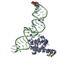 1dulS S: Starting model for refinement C: citing same article ( |
|---|---|
| Similar structure data |

-Links
 PDBj
PDBj


- Assembly
| Deposited unit | 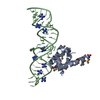
| ||||||||
|---|---|---|---|---|---|---|---|---|---|
| 1 |
| ||||||||
| Unit cell |
|
-Components
| #1: RNA chain | Mass: 15885.515 Da / Num. of mol.: 1 / Fragment: DOMAIN IV / Mutation: C132U, U133G, A175U Source method: isolated from a genetically manipulated source | ||
|---|---|---|---|
| #2: Protein | Mass: 12336.646 Da / Num. of mol.: 1 / Fragment: C TERMINAL DOMAIN (RESIDUES 328-432) Source method: isolated from a genetically manipulated source Source: (gene. exp.) | ||
| #3: Chemical | ChemComp-NCO /   Mass: 161.116 Da / Num. of mol.: 8 / Source method: obtained synthetically / Formula: CoH18N6 Mass: 161.116 Da / Num. of mol.: 8 / Source method: obtained synthetically / Formula: CoH18N6Has protein modification | Y | |
-Experimental details
-Experiment
| Experiment | Method: X-RAY DIFFRACTION / Number of used crystals: 1 |
|---|
- Sample preparation
| Crystal | Density Matthews: 2.94 Å3/Da / Density % sol: 58.1 % | ||||||||||||||||||||||||||||||||
|---|---|---|---|---|---|---|---|---|---|---|---|---|---|---|---|---|---|---|---|---|---|---|---|---|---|---|---|---|---|---|---|---|---|
| Crystal grow | Temperature: 293 K / Method: vapor diffusion, sitting drop / pH: 5.6 Details: 20Mm NaOH-MES pH 5.6, 200mM KCl, 10% Isopropanol, 5mM Cobalt Hexamine, VAPOR DIFFUSION, SITTING DROP, temperature 293K | ||||||||||||||||||||||||||||||||
| Components of the solutions |
|
-Data collection
| Diffraction |
| |||||||||
|---|---|---|---|---|---|---|---|---|---|---|
| Diffraction source | Source: ROTATING ANODE / Type: RIGAKU / Wavelength: 1.5418 Å | |||||||||
| Detector | Type: RIGAKU RAXIS / Detector: IMAGE PLATE / Date: Feb 18, 2006 | |||||||||
| Radiation | Protocol: SINGLE WAVELENGTH / Monochromatic (M) / Laue (L): M / Scattering type: x-ray | |||||||||
| Radiation wavelength | Wavelength: 1.5418 Å / Relative weight: 1 | |||||||||
| Reflection | Resolution: 2.5→39.34 Å / Num. obs: 10959 / % possible obs: 96.4 % / Redundancy: 7.72 % / Rmerge(I) obs: 0.065 / Χ2: 1.01 / Net I/σ(I): 18.7 / Scaling rejects: 641 | |||||||||
| Reflection shell | Resolution: 2.5→2.59 Å / Redundancy: 7.59 % / Rmerge(I) obs: 0.348 / Mean I/σ(I) obs: 4.8 / Num. measured all: 8128 / Num. unique all: 1071 / Χ2: 0.72 / % possible all: 96.1 |
-Phasing
| Phasing MR | Cor.coef. Fo:Fc: 0.86 / Packing: 0.392
|
|---|
- Processing
| Software |
| ||||||||||||||||||||||||||||
|---|---|---|---|---|---|---|---|---|---|---|---|---|---|---|---|---|---|---|---|---|---|---|---|---|---|---|---|---|---|
| Refinement | Method to determine structure: MOLECULAR REPLACEMENT Starting model: PDB ENTRY 1DUL Resolution: 2.5→50 Å / Cross valid method: THROUGHOUT / σ(F): 0
| ||||||||||||||||||||||||||||
| Solvent computation | Bsol: 45 Å2 | ||||||||||||||||||||||||||||
| Displacement parameters | Biso mean: 56.38 Å2
| ||||||||||||||||||||||||||||
| Refinement step | Cycle: LAST / Resolution: 2.5→50 Å
| ||||||||||||||||||||||||||||
| Xplor file |
|
