 Movie
Movie Controller

[English] 日本語
 Yorodumi
- PDB-2pv3: Crystallographic Structure of SurA fragment lacking the second pe... -
+ Open data

- Basic information
| Entry | Database: PDB / ID: 2pv3 | ||||||
|---|---|---|---|---|---|---|---|
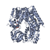
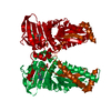
| Title | Crystallographic Structure of SurA fragment lacking the second peptidyl-prolyl isomerase domain complexed with peptide NFTLKFWDIFRK | ||||||
 Components Components |
| ||||||
 Keywords Keywords | ISOMERASE / Survival protein A / Peptidyl-prolyl cis-trans isomerase domain | ||||||
| Function / homology |  Function and homology information Function and homology information: / Gram-negative-bacterium-type cell outer membrane assembly / Secretion of toxins / peptide binding / peptidylprolyl isomerase / peptidyl-prolyl cis-trans isomerase activity / : / outer membrane-bounded periplasmic space / protein folding / protein stabilization Similarity search - Function | ||||||
| Biological species |  | ||||||
| Method |  X-RAY DIFFRACTION / SYNCHROTRON / MAD / Resolution: 3.39 Å X-RAY DIFFRACTION / SYNCHROTRON / MAD / Resolution: 3.39 Å | ||||||
 Authors Authors | Xu, X. / McKay, D.B. | ||||||
 Citation Citation | Journal: J.Mol.Biol. / Year: 2007 Title: The Periplasmic Bacterial Molecular Chaperone SurA Adapts its Structure to Bind Peptides in Different Conformations to Assert a Sequence Preference for Aromatic Residues. Authors: Xu, X. / Wang, S. / Hu, Y.X. / McKay, D.B. #1: Journal: Structure / Year: 2002Title: Crystallographic structure of SurA, a molecular chaperone that facilitates folding of outer membrane porins Authors: Bitto, E. / McKay, D.B. | ||||||
| History |
|
- Structure visualization
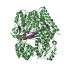
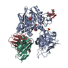
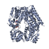
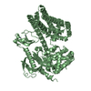
| Structure viewer | Molecule: MolmilJmol/JSmol |
|---|
- Downloads & links
-Download
| PDBx/mmCIF format | 2pv3.cif.gz | 116.1 KB | Display | PDBx/mmCIF format |
|---|---|---|---|---|
| PDB format | pdb2pv3.ent.gz | 93 KB | Display | PDB format |
| PDBx/mmJSON format | 2pv3.json.gz | Tree view | PDBx/mmJSON format | |
| Others |  Other downloads Other downloads |
-Validation report
| Arichive directory | https://data.pdbj.org/pub/pdb/validation_reports/pv/2pv3ftp://data.pdbj.org/pub/pdb/validation_reports/pv/2pv3 | HTTPS FTP |
|---|
-Related structure data
| Related structure data |  2pv1C  2pv2C  1m5yS S: Starting model for refinement C: citing same article ( |
|---|---|
| Similar structure data |


-Links
 PDBj
PDBj

- Assembly
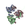
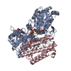
| Deposited unit | 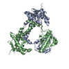
| ||||||||
|---|---|---|---|---|---|---|---|---|---|
| 1 |
| ||||||||
| Unit cell |
|
-Components
| #1: Protein | Mass: 33221.621 Da / Num. of mol.: 2 Fragment: Survivial protein A fragment from which the second peptidyl-prolyl isomerase domain has been deleted Source method: isolated from a genetically manipulated source Source: (gene. exp.) #2: Protein/peptide | | Mass: 1617.910 Da / Num. of mol.: 1 / Source method: obtained synthetically |
|---|
-Experimental details
-Experiment
| Experiment | Method: X-RAY DIFFRACTION / Number of used crystals: 1 |
|---|
- Sample preparation
| Crystal grow | Temperature: 291 K / Method: vapor diffusion, hanging drop / pH: 6.5 Details: 1.4~1.8M sodium chloride, 0.1M potassium dihydrogen phosphate, 0.1 M sodium dihydrogen phosphate, 0.1M MES buffer, pH6.5, VAPOR DIFFUSION, HANGING DROP, temperature 291K |
|---|
-Data collection
| Diffraction |
| ||||||||||||||||||
|---|---|---|---|---|---|---|---|---|---|---|---|---|---|---|---|---|---|---|---|
| Diffraction source |
| ||||||||||||||||||
| Detector | Type: MARMOSAIC 325 mm CCD / Detector: CCD / Date: Jun 21, 2006 | ||||||||||||||||||
| Radiation | Protocol: MAD / Monochromatic (M) / Laue (L): M / Scattering type: x-ray | ||||||||||||||||||
| Radiation wavelength |
| ||||||||||||||||||
| Reflection | Resolution: 3.39→50 Å / Num. obs: 33486 / % possible obs: 99.1 % / Redundancy: 4.2 % / Rsym value: 0.074 / Net I/σ(I): 17.5 | ||||||||||||||||||
| Reflection shell | Resolution: 3.39→3.52 Å / Redundancy: 3.5 % / Num. unique all: 3326 / Rsym value: 0.422 / % possible all: 99.8 |

- Processing
| Software |
| ||||||||||||||||||||||||||||||||||||||||||||||||||||||||||||||||||||||||||||||||||||||||||||||||||||
|---|---|---|---|---|---|---|---|---|---|---|---|---|---|---|---|---|---|---|---|---|---|---|---|---|---|---|---|---|---|---|---|---|---|---|---|---|---|---|---|---|---|---|---|---|---|---|---|---|---|---|---|---|---|---|---|---|---|---|---|---|---|---|---|---|---|---|---|---|---|---|---|---|---|---|---|---|---|---|---|---|---|---|---|---|---|---|---|---|---|---|---|---|---|---|---|---|---|---|---|---|---|
| Refinement | Method to determine structure: MAD Starting model: PDB ENTRY 1M5Y Resolution: 3.39→50 Å / Cor.coef. Fo:Fc: 0.923 / Cor.coef. Fo:Fc free: 0.908 / SU B: 22.754 / SU ML: 0.328 / Cross valid method: THROUGHOUT / σ(F): 0 / ESU R: 0.501 / ESU R Free: 0.396 / Stereochemistry target values: MAXIMUM LIKELIHOOD / Details: HYDROGENS HAVE BEEN ADDED IN THE RIDING POSITIONS
| ||||||||||||||||||||||||||||||||||||||||||||||||||||||||||||||||||||||||||||||||||||||||||||||||||||
| Solvent computation | Ion probe radii: 0.8 Å / Shrinkage radii: 0.8 Å / VDW probe radii: 1.2 Å / Solvent model: MASK | ||||||||||||||||||||||||||||||||||||||||||||||||||||||||||||||||||||||||||||||||||||||||||||||||||||
| Displacement parameters | Biso mean: 171.417 Å2
| ||||||||||||||||||||||||||||||||||||||||||||||||||||||||||||||||||||||||||||||||||||||||||||||||||||
| Refine analyze |
| ||||||||||||||||||||||||||||||||||||||||||||||||||||||||||||||||||||||||||||||||||||||||||||||||||||
| Refinement step | Cycle: LAST / Resolution: 3.39→50 Å
| ||||||||||||||||||||||||||||||||||||||||||||||||||||||||||||||||||||||||||||||||||||||||||||||||||||
| Refine LS restraints |
| ||||||||||||||||||||||||||||||||||||||||||||||||||||||||||||||||||||||||||||||||||||||||||||||||||||
| LS refinement shell | Resolution: 3.39→3.478 Å / Total num. of bins used: 20
|
