 Movie
Movie Controller

[English] 日本語
 Yorodumi
- PDB-2ptw: Crystal Structure of the T. brucei enolase complexed with sulphat... -
+ Open data

- Basic information
| Entry | Database: PDB / ID: 2ptw | ||||||
|---|---|---|---|---|---|---|---|
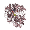
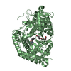
| Title | Crystal Structure of the T. brucei enolase complexed with sulphate, identification of a metal binding site IV | ||||||
 Components Components | Enolase | ||||||
 Keywords Keywords | LYASE / GLYCOLYSIS / HIS-TAG | ||||||
| Function / homology |  Function and homology information Function and homology informationphosphopyruvate hydratase / phosphopyruvate hydratase complex / phosphopyruvate hydratase activity / ciliary plasm / glycolytic process / magnesium ion binding / cytoplasm / cytosol Similarity search - Function | ||||||
| Biological species |  | ||||||
| Method |  X-RAY DIFFRACTION / MOLECULAR REPLACEMENT / Resolution: 1.9 Å X-RAY DIFFRACTION / MOLECULAR REPLACEMENT / Resolution: 1.9 Å | ||||||
 Authors Authors | Navarro, M.V.A.S. / Rigden, D.J. / Garratt, R.C. / Dias, S.M.G. | ||||||
 Citation Citation | Journal: Febs J. / Year: 2007 Title: Structural flexibility in Trypanosoma brucei enolase revealed by X-ray crystallography and molecular dynamics. Authors: Navarro, M.V. / Gomes Dias, S.M. / Mello, L.V. / da Silva Giotto, M.T. / Gavalda, S. / Blonski, C. / Garratt, R.C. / Rigden, D.J. #1: Journal: J.Mol.Biol. / Year: 2003Title: The Crystal Structure of Trypanosoma Brucei Enolase: Visualisation of the Inhibitory Metal Binding Site III and Potential as Target for Selective, Irreversible Inhibition Authors: da Silva Giotto, M.T. / Hannaert, V. / Vertommen, D. / Navarro, M.V.A.S. / Rider, M.H. / Michels, P.A.M. / Garratt, R.C. / Rigden, D.J. | ||||||
| History |
|
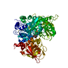
- Structure visualization
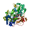
| Structure viewer | Molecule: MolmilJmol/JSmol |
|---|
- Downloads & links
-Download
| PDBx/mmCIF format | 2ptw.cif.gz | 186.5 KB | Display | PDBx/mmCIF format |
|---|---|---|---|---|
| PDB format | pdb2ptw.ent.gz | 146.6 KB | Display | PDB format |
| PDBx/mmJSON format | 2ptw.json.gz | Tree view | PDBx/mmJSON format | |
| Others |  Other downloads Other downloads |
-Validation report
| Summary document | 2ptw_validation.pdf.gz | 451.6 KB | Display | wwPDB validaton report |
|---|---|---|---|---|
| Full document | 2ptw_full_validation.pdf.gz | 457.6 KB | Display | |
| Data in XML | 2ptw_validation.xml.gz | 20.2 KB | Display | |
| Data in CIF | 2ptw_validation.cif.gz | 30.1 KB | Display | |
| Arichive directory | https://data.pdbj.org/pub/pdb/validation_reports/pt/2ptwftp://data.pdbj.org/pub/pdb/validation_reports/pt/2ptw | HTTPS FTP |
-Related structure data
| Related structure data | 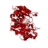 2ptxC  2ptyC  2ptzC 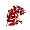 2pu0C 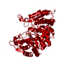 2pu1C 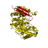 1oepS S: Starting model for refinement C: citing same article ( |
|---|---|
| Similar structure data |

-Links
 PDBj
PDBj
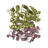
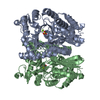
- Assembly
| Deposited unit | 
| ||||||||
|---|---|---|---|---|---|---|---|---|---|
| 1 | 
| ||||||||
| Unit cell |
| ||||||||
| Components on special symmetry positions |
| ||||||||
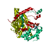
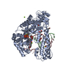
| Details | The biological assembly is a dimer generated from the monomer in the asymmetric unit by the operations: -x, y, -z+1/2 |
-Components
| #1: Protein | Mass: 46904.434 Da / Num. of mol.: 1 / Mutation: R28K Source method: isolated from a genetically manipulated source Source: (gene. exp.)  | ||||||
|---|---|---|---|---|---|---|---|
| #2: Chemical |   Mass: 65.409 Da / Num. of mol.: 2 / Source method: obtained synthetically / Formula: Zn Mass: 65.409 Da / Num. of mol.: 2 / Source method: obtained synthetically / Formula: Zn#3: Chemical | ChemComp-SO4 / |   Mass: 96.063 Da / Num. of mol.: 1 / Source method: obtained synthetically / Formula: SO4 Mass: 96.063 Da / Num. of mol.: 1 / Source method: obtained synthetically / Formula: SO4#4: Chemical |   Mass: 62.068 Da / Num. of mol.: 3 / Source method: obtained synthetically / Formula: C2H6O2 Mass: 62.068 Da / Num. of mol.: 3 / Source method: obtained synthetically / Formula: C2H6O2#5: Water | ChemComp-HOH / |  Mass: 18.015 Da / Num. of mol.: 308 / Source method: isolated from a natural source / Formula: H2O Mass: 18.015 Da / Num. of mol.: 308 / Source method: isolated from a natural source / Formula: H2O |
-Experimental details
-Experiment
| Experiment | Method: X-RAY DIFFRACTION / Number of used crystals: 1 |
|---|
- Sample preparation
| Crystal | Density Matthews: 2.4 Å3/Da / Density % sol: 48.74 % |
|---|---|
| Crystal grow | Temperature: 291 K / Method: vapor diffusion, hanging drop / pH: 6.5 Details: 10 % (w/v) PEG1000, 0.01 M ZnSO4 or ZnCl2, and 0.1 M MES, pH 6.5, VAPOR DIFFUSION, HANGING DROP, temperature 291K |
-Data collection
| Diffraction | Mean temperature: 100 K |
|---|---|
| Diffraction source | Source: ROTATING ANODE / Type: RIGAKU ULTRAX 18 / Wavelength: 1.54 Å |
| Detector | Type: MAR scanner 345 mm plate / Detector: IMAGE PLATE / Date: Oct 4, 2004 / Details: MIRRORS |
| Radiation | Monochromator: GRAPHITE / Protocol: SINGLE WAVELENGTH / Monochromatic (M) / Laue (L): M / Scattering type: x-ray |
| Radiation wavelength | Wavelength: 1.54 Å / Relative weight: 1 |
| Reflection | Resolution: 1.9→26 Å / Num. all: 36064 / Num. obs: 35703 / % possible obs: 99 % / Observed criterion σ(F): 1 / Observed criterion σ(I): 1 / Redundancy: 3.2 % / Biso Wilson estimate: 23.4 Å2 / Rmerge(I) obs: 0.058 / Net I/σ(I): 17 |
| Reflection shell | Resolution: 1.9→2 Å / Redundancy: 3.2 % / Rmerge(I) obs: 0.542 / Mean I/σ(I) obs: 3.9 / Num. unique all: 5081 / % possible all: 96.7 |
- Processing
| Software |
| ||||||||||||||||||||||||||||||||||||||||||||||||||||||||||||||||||||||||||||||||||||||||||
|---|---|---|---|---|---|---|---|---|---|---|---|---|---|---|---|---|---|---|---|---|---|---|---|---|---|---|---|---|---|---|---|---|---|---|---|---|---|---|---|---|---|---|---|---|---|---|---|---|---|---|---|---|---|---|---|---|---|---|---|---|---|---|---|---|---|---|---|---|---|---|---|---|---|---|---|---|---|---|---|---|---|---|---|---|---|---|---|---|---|---|---|
| Refinement | Method to determine structure: MOLECULAR REPLACEMENT Starting model: 1OEP Resolution: 1.9→25.98 Å / Cor.coef. Fo:Fc: 0.939 / Cor.coef. Fo:Fc free: 0.922 / SU B: 10.84 / SU ML: 0.14 / Isotropic thermal model: Isotropic / Cross valid method: THROUGHOUT / σ(F): 0 / σ(I): 0 / ESU R: 0.17 / ESU R Free: 0.153 / Stereochemistry target values: MAXIMUM LIKELIHOOD / Details: HYDROGENS HAVE BEEN ADDED IN THE RIDING POSITIONS
| ||||||||||||||||||||||||||||||||||||||||||||||||||||||||||||||||||||||||||||||||||||||||||
| Solvent computation | Ion probe radii: 0.8 Å / Shrinkage radii: 0.8 Å / VDW probe radii: 1.4 Å / Solvent model: MASK | ||||||||||||||||||||||||||||||||||||||||||||||||||||||||||||||||||||||||||||||||||||||||||
| Displacement parameters | Biso mean: 20.502 Å2
| ||||||||||||||||||||||||||||||||||||||||||||||||||||||||||||||||||||||||||||||||||||||||||
| Refinement step | Cycle: LAST / Resolution: 1.9→25.98 Å
| ||||||||||||||||||||||||||||||||||||||||||||||||||||||||||||||||||||||||||||||||||||||||||
| Refine LS restraints |
| ||||||||||||||||||||||||||||||||||||||||||||||||||||||||||||||||||||||||||||||||||||||||||
| LS refinement shell | Resolution: 1.9→2.002 Å / Total num. of bins used: 10
| ||||||||||||||||||||||||||||||||||||||||||||||||||||||||||||||||||||||||||||||||||||||||||
| Refinement TLS params. | Method: refined / Refine-ID: X-RAY DIFFRACTION
| ||||||||||||||||||||||||||||||||||||||||||||||||||||||||||||||||||||||||||||||||||||||||||
| Refinement TLS group |
|
