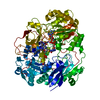
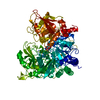
 Movie
Movie Controller


+ Open data

- Basic information
| Entry | Database: PDB / ID: 1.0E+60 | ||||||
|---|---|---|---|---|---|---|---|
| Title | OXIDIZED DMSO REDUCTASE EXPOSED TO HEPES - Structure II BUFFER | ||||||
 Components Components | Dimethyl sulfoxide/trimethylamine N-oxide reductase | ||||||
 Keywords Keywords | OXIDOREDUCTASE / REDUCTASE / DMSO / MOLYBDOPTERIN | ||||||
| Function / homology |  Function and homology information Function and homology informationrespiratory dimethylsulfoxide reductase / trimethylamine-N-oxide reductase / trimethylamine-N-oxide reductase (cytochrome c) activity / molybdenum ion binding / molybdopterin cofactor binding / anaerobic respiration / outer membrane-bounded periplasmic space / electron transfer activity Similarity search - Function | ||||||
| Biological species |  Rhodobacter capsulatus (bacteria) Rhodobacter capsulatus (bacteria) | ||||||
| Method |  X-RAY DIFFRACTION / SYNCHROTRON / MOLECULAR REPLACEMENT / Resolution: 2 Å X-RAY DIFFRACTION / SYNCHROTRON / MOLECULAR REPLACEMENT / Resolution: 2 Å | ||||||
 Authors Authors | Bailey, S. / Bennett, B. / Adams, B. / Smith, A.T. / Bray, R.C. | ||||||
 Citation Citation | Journal: Biochemistry / Year: 2000 Title: Reversible Dissociation of Thiolate Ligands from Molybdenum in an Enzyme of the Dimethyl Sulfoxide Reductase Family Authors: Bray, R.C. / Adams, B. / Smith, A.T. / Bennett, B. / Bailey, S. | ||||||
| History |
|
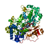
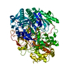
- Structure visualization
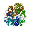
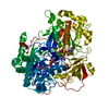
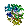
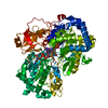
| Structure viewer | Molecule: MolmilJmol/JSmol |
|---|
- Downloads & links
-Download
| PDBx/mmCIF format | 1e60.cif.gz | 341.2 KB | Display | PDBx/mmCIF format |
|---|---|---|---|---|
| PDB format | pdb1e60.ent.gz | 270.7 KB | Display | PDB format |
| PDBx/mmJSON format | 1e60.json.gz | Tree view | PDBx/mmJSON format | |
| Others |  Other downloads Other downloads |
-Validation report
| Arichive directory | https://data.pdbj.org/pub/pdb/validation_reports/e6/1e60ftp://data.pdbj.org/pub/pdb/validation_reports/e6/1e60 | HTTPS FTP |
|---|
-Related structure data


-Links
 PDBj
PDBj


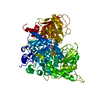
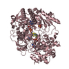
- Assembly
| Deposited unit | 
| ||||||||
|---|---|---|---|---|---|---|---|---|---|
| 1 | 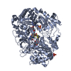
| ||||||||
| 2 | 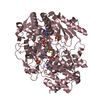
| ||||||||
| Unit cell |
| ||||||||
| Noncrystallographic symmetry (NCS) | NCS oper: (Code: given Matrix: (0.03048, -0.07809, -0.99648), Vector: |
-Components
| #1: Protein | Mass: 89525.938 Da / Num. of mol.: 2 / Source method: isolated from a natural source / Source: (natural) Rhodobacter capsulatus (bacteria) / Cellular location: PERIPLASMReferences: UniProt: Q52675, trimethylamine-N-oxide reductase, respiratory dimethylsulfoxide reductase #2: Chemical | ChemComp-PGD /   Mass: 738.541 Da / Num. of mol.: 4 / Source method: obtained synthetically / Formula: C20H24N10O13P2S2 Mass: 738.541 Da / Num. of mol.: 4 / Source method: obtained synthetically / Formula: C20H24N10O13P2S2#3: Chemical |   Mass: 127.939 Da / Num. of mol.: 2 / Source method: obtained synthetically / Formula: MoO2 Mass: 127.939 Da / Num. of mol.: 2 / Source method: obtained synthetically / Formula: MoO2#4: Chemical | ChemComp-SO4 /   Mass: 96.063 Da / Num. of mol.: 4 / Source method: obtained synthetically / Formula: SO4 Mass: 96.063 Da / Num. of mol.: 4 / Source method: obtained synthetically / Formula: SO4#5: Water | ChemComp-HOH / |  Mass: 18.015 Da / Num. of mol.: 1318 / Source method: isolated from a natural source / Formula: H2O Mass: 18.015 Da / Num. of mol.: 1318 / Source method: isolated from a natural source / Formula: H2OCompound details | REDUCES VARIOUS N-OXIDE AND SULFOXIDE COMPOUNDS INCLUDING TRIMETHYLAMINE N-OXIDE DURING ANAEROBIC ...REDUCES VARIOUS N-OXIDE AND SULFOXIDE COMPOUNDS INCLUDING TRIMETHYLA | Has protein modification | N | |
|---|
-Experimental details
-Experiment
| Experiment | Method: X-RAY DIFFRACTION / Number of used crystals: 1 |
|---|
- Sample preparation
| Crystal | Density Matthews: 2.55 Å3/Da / Density % sol: 45 % | ||||||||||||||||||||||||||||||
|---|---|---|---|---|---|---|---|---|---|---|---|---|---|---|---|---|---|---|---|---|---|---|---|---|---|---|---|---|---|---|---|
| Crystal grow | pH: 7.5 Details: 0.1M HEPES BUFFER, PH 7.5 2M AMMONIUM SULPHATE 3-4% PEG 400 | ||||||||||||||||||||||||||||||
| Crystal grow | *PLUS Temperature: 20-22 ℃ / pH: 7 / Method: vapor diffusion | ||||||||||||||||||||||||||||||
| Components of the solutions | *PLUS
|
-Data collection
| Diffraction | Mean temperature: 100 K |
|---|---|
| Diffraction source | Source: SYNCHROTRON / Site: ESRF  / Beamline: BM14 / Wavelength: 0.97 / Beamline: BM14 / Wavelength: 0.97 |
| Detector | Type: MARRESEARCH / Detector: IMAGE PLATE / Date: Nov 15, 1998 |
| Radiation | Protocol: SINGLE WAVELENGTH / Monochromatic (M) / Laue (L): M / Scattering type: x-ray |
| Radiation wavelength | Wavelength: 0.97 Å / Relative weight: 1 |
| Reflection | Resolution: 2→20 Å / Num. obs: 123010 / % possible obs: 98.1 % / Observed criterion σ(I): 0 / Redundancy: 3.8 % / Biso Wilson estimate: 17 Å2 / Rmerge(I) obs: 0.083 / Net I/σ(I): 5.6 |
| Reflection shell | Mean I/σ(I) obs: 5.1 |
| Reflection | *PLUS Num. measured all: 465630 |
- Processing
| Software |
| ||||||||||||||||||||||||||||||||||||||||||||||||||||||||||||||||||||||||||||||||||||
|---|---|---|---|---|---|---|---|---|---|---|---|---|---|---|---|---|---|---|---|---|---|---|---|---|---|---|---|---|---|---|---|---|---|---|---|---|---|---|---|---|---|---|---|---|---|---|---|---|---|---|---|---|---|---|---|---|---|---|---|---|---|---|---|---|---|---|---|---|---|---|---|---|---|---|---|---|---|---|---|---|---|---|---|---|---|
| Refinement | Method to determine structure: MOLECULAR REPLACEMENT Starting model: DMSO REDUCTASE FROM RHODOBACTER CAPSULATUS Resolution: 2→20 Å / SU B: 4.2 / SU ML: 0.12 / Cross valid method: THROUGHOUT / σ(F): 0 / ESU R: 0.18 / ESU R Free: 0.16
| ||||||||||||||||||||||||||||||||||||||||||||||||||||||||||||||||||||||||||||||||||||
| Refinement step | Cycle: LAST / Resolution: 2→20 Å
| ||||||||||||||||||||||||||||||||||||||||||||||||||||||||||||||||||||||||||||||||||||
| Refine LS restraints |
| ||||||||||||||||||||||||||||||||||||||||||||||||||||||||||||||||||||||||||||||||||||
| Software | *PLUS Name: REFMAC / Classification: refinement | ||||||||||||||||||||||||||||||||||||||||||||||||||||||||||||||||||||||||||||||||||||
| Refinement | *PLUS Lowest resolution: 30 Å / Rfactor obs: 0.194 | ||||||||||||||||||||||||||||||||||||||||||||||||||||||||||||||||||||||||||||||||||||
| Solvent computation | *PLUS | ||||||||||||||||||||||||||||||||||||||||||||||||||||||||||||||||||||||||||||||||||||
| Displacement parameters | *PLUS |
