 Movie
Movie Controller

[English] 日本語
 Yorodumi
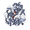
- PDB-4dmr: REDUCED DMSO REDUCTASE FROM RHODOBACTER CAPSULATUS WITH BOUND DMS... -
+ Open data

- Basic information
| Entry | Database: PDB / ID: 4dmr | ||||||
|---|---|---|---|---|---|---|---|
| Title | REDUCED DMSO REDUCTASE FROM RHODOBACTER CAPSULATUS WITH BOUND DMSO SUBSTRATE | ||||||
 Components Components | DMSO REDUCTASE | ||||||
 Keywords Keywords | OXIDOREDUCTASE / DMSO / MOLYBDOPTERIN / SUBSTRATE BOUND | ||||||
| Function / homology |  Function and homology information Function and homology informationrespiratory dimethylsulfoxide reductase / trimethylamine-N-oxide reductase / trimethylamine-N-oxide reductase (cytochrome c) activity / molybdenum ion binding / molybdopterin cofactor binding / anaerobic respiration / outer membrane-bounded periplasmic space / electron transfer activity Similarity search - Function | ||||||
| Biological species |  Rhodobacter capsulatus (bacteria) Rhodobacter capsulatus (bacteria) | ||||||
| Method |  X-RAY DIFFRACTION / SYNCHROTRON / DIFFERENCE MAPS USING OXIDISED STRUCTURE / Resolution: 1.9 Å X-RAY DIFFRACTION / SYNCHROTRON / DIFFERENCE MAPS USING OXIDISED STRUCTURE / Resolution: 1.9 Å | ||||||
 Authors Authors | Mcalpine, A.S. / Bailey, S. | ||||||
 Citation Citation | Journal: J.Mol.Biol. / Year: 1998 Title: The high resolution crystal structure of DMSO reductase in complex with DMSO. Authors: McAlpine, A.S. / McEwan, A.G. / Bailey, S. #1: Journal: J.Biol.Inorg.Chem. / Year: 1997Title: Molybdenum Active Centre of Dmso Reductase from Rhodobacter Capsulatus: Crystal Structure of the Oxidised Enzyme at 1.82-A Resolution and the Dithionite-Reduced Enzyme at 2.8-A Resolution Authors: Mcalpine, A.S. / Mcewan, A.G. / Shaw, A. / Bailey, S. #2: Journal: Acta Crystallogr.,Sect.D / Year: 1996Title: Preliminary Crystallographic Studies of Dimethylsulfoxide Reductase from Rhodobacter Capsulatus Authors: Bailey, S. / Mcalpine, A.S. / Duke, E.M.H. / Benson, N. / Mcewan, A.G. | ||||||
| History |
|
- Structure visualization
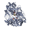
| Structure viewer | Molecule: MolmilJmol/JSmol |
|---|
- Downloads & links
-Download
| PDBx/mmCIF format | 4dmr.cif.gz | 177 KB | Display | PDBx/mmCIF format |
|---|---|---|---|---|
| PDB format | pdb4dmr.ent.gz | 134.5 KB | Display | PDB format |
| PDBx/mmJSON format | 4dmr.json.gz | Tree view | PDBx/mmJSON format | |
| Others |  Other downloads Other downloads |
-Validation report
| Arichive directory | https://data.pdbj.org/pub/pdb/validation_reports/dm/4dmrftp://data.pdbj.org/pub/pdb/validation_reports/dm/4dmr | HTTPS FTP |
|---|
-Related structure data
| Similar structure data |
|---|


-Links
 PDBj
PDBj


- Assembly
| Deposited unit | 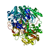
| ||||||||
|---|---|---|---|---|---|---|---|---|---|
| 1 |
| ||||||||
| Unit cell |
|
-Components
-Protein , 1 types, 1 molecules A
| #1: Protein | Mass: 89482.914 Da / Num. of mol.: 1 / Source method: isolated from a natural source / Source: (natural) Rhodobacter capsulatus (bacteria) / Cellular location: PERIPLASM / Strain: H123 / References: UniProt: Q52675 |
|---|
-Non-polymers , 5 types, 479 molecules 


| #2: Chemical |  Mass: 738.541 Da / Num. of mol.: 2 / Source method: obtained synthetically / Formula: C20H24N10O13P2S2 Mass: 738.541 Da / Num. of mol.: 2 / Source method: obtained synthetically / Formula: C20H24N10O13P2S2#3: Chemical | ChemComp-4MO / |  Mass: 95.940 Da / Num. of mol.: 1 / Source method: obtained synthetically / Formula: Mo Mass: 95.940 Da / Num. of mol.: 1 / Source method: obtained synthetically / Formula: Mo#4: Chemical | ChemComp-O / |  Mass: 15.999 Da / Num. of mol.: 1 / Source method: obtained synthetically / Formula: O Mass: 15.999 Da / Num. of mol.: 1 / Source method: obtained synthetically / Formula: O#5: Chemical | ChemComp-DMS / |  Mass: 78.133 Da / Num. of mol.: 1 / Source method: obtained synthetically / Formula: C2H6OS / Comment: DMSO, precipitant*YM Mass: 78.133 Da / Num. of mol.: 1 / Source method: obtained synthetically / Formula: C2H6OS / Comment: DMSO, precipitant*YM#6: Water | ChemComp-HOH / | Mass: 18.015 Da / Num. of mol.: 474 / Source method: isolated from a natural source / Formula: H2O |
|---|
-Details
| Nonpolymer details | STRUCTURE HAS DMSO (SUBSTRATE) BOUND AT THE ACTIVE SITE. THE ACTIVE SITE IS IN THE MO(IV) REDUCED STATE. |
|---|
-Experimental details
-Experiment
| Experiment | Method: X-RAY DIFFRACTION / Number of used crystals: 1 |
|---|
- Sample preparation
| Crystal | Density Matthews: 2.11 Å3/Da / Density % sol: 35.1 % | ||||||||||||||||||||||||||||||
|---|---|---|---|---|---|---|---|---|---|---|---|---|---|---|---|---|---|---|---|---|---|---|---|---|---|---|---|---|---|---|---|
| Crystal grow | pH: 5.5 Details: 0.1M CITRATE BUFFER, PH 4.8 - 5.2 20 - 3% PEG 4000 20% ETHANOL, pH 5.5 A FEW UL OF NEAT DMS (DIMETHYL SULFIDE) WAS ADDED TO THE DROP CONTAINING THE CRYSTAL IMMEDIATELY BEFORE DATA COLLECTION. PH range: 4.8-5.2 | ||||||||||||||||||||||||||||||
| Crystal | *PLUS | ||||||||||||||||||||||||||||||
| Crystal grow | *PLUS Method: vapor diffusionDetails: Bailey, S., (1996) Acta Crystallogr.,Sect.D, 52, 194. PH range low: 5.6 / PH range high: 5 | ||||||||||||||||||||||||||||||
| Components of the solutions | *PLUS
|
-Data collection
| Diffraction | Mean temperature: 277 K |
|---|---|
| Diffraction source | Source: SYNCHROTRON / Site: SRS  / Beamline: PX9.6 / Wavelength: 0.87 / Beamline: PX9.6 / Wavelength: 0.87 |
| Detector | Type: MARRESEARCH / Detector: IMAGE PLATE / Date: Oct 3, 1996 / Details: MIRRORS |
| Radiation | Monochromator: SI(111) / Monochromatic (M) / Laue (L): M / Scattering type: x-ray |
| Radiation wavelength | Wavelength: 0.87 Å / Relative weight: 1 |
| Reflection | Resolution: 1.9→17.3 Å / Num. obs: 58725 / % possible obs: 96.6 % / Observed criterion σ(I): 0 / Redundancy: 3.9 % / Rmerge(I) obs: 0.096 / Net I/σ(I): 6 |
| Reflection shell | Resolution: 1.9→2.03 Å / Redundancy: 3.8 % / Rmerge(I) obs: 0.282 / Mean I/σ(I) obs: 2.3 / % possible all: 96.6 |
| Reflection | *PLUS Num. measured all: 227149 |
| Reflection shell | *PLUS % possible obs: 90.5 % |
- Processing
| Software |
| ||||||||||||||||||||
|---|---|---|---|---|---|---|---|---|---|---|---|---|---|---|---|---|---|---|---|---|---|
| Refinement | Method to determine structure: DIFFERENCE MAPS USING OXIDISED STRUCTURE Starting model: OXIDISED STRUCTURE FOR DIFFERENCES IN DIFFERENCE MAP Resolution: 1.9→20 Å / Cross valid method: USE OF SINGLE FREE R SET / σ(F): 0
| ||||||||||||||||||||
| Refinement step | Cycle: LAST / Resolution: 1.9→20 Å
| ||||||||||||||||||||
| Software | *PLUS Name: REFMAC / Classification: refinement | ||||||||||||||||||||
| Refinement | *PLUS Num. reflection all: 58725 / Rfactor obs: 0.16 | ||||||||||||||||||||
| Solvent computation | *PLUS | ||||||||||||||||||||
| Displacement parameters | *PLUS | ||||||||||||||||||||
| Refine LS restraints | *PLUS
|
