 Movie
Movie Controller

[English] 日本語
 Yorodumi
- PDB-1oep: Structure of Trypanosoma brucei enolase reveals the inhibitory di... -
+ Open data

- Basic information
| Entry | Database: PDB / ID: 1oep | ||||||
|---|---|---|---|---|---|---|---|
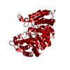
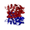
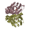
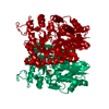
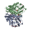
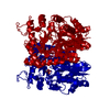
| Title | Structure of Trypanosoma brucei enolase reveals the inhibitory divalent metal site | ||||||
 Components Components | ENOLASE | ||||||
 Keywords Keywords | LYASE / GLYCOLYSIS / HIS-TAG | ||||||
| Function / homology |  Function and homology information Function and homology informationphosphopyruvate hydratase / phosphopyruvate hydratase complex / phosphopyruvate hydratase activity / ciliary plasm / glycolytic process / magnesium ion binding / cytoplasm / cytosol Similarity search - Function | ||||||
| Biological species |  | ||||||
| Method |  X-RAY DIFFRACTION / MOLECULAR REPLACEMENT / Resolution: 2.3 Å X-RAY DIFFRACTION / MOLECULAR REPLACEMENT / Resolution: 2.3 Å | ||||||
 Authors Authors | Da Silva giotto, M.T. / Navarro, M.V.A.S. / Garratt, R.C. / Rigden, D.J. | ||||||
 Citation Citation | Journal: J.Mol.Biol. / Year: 2003 Title: The Crystal Structure of Trypanosoma Brucei Enolase: Visualisation of the Inhibitory Metal Binding Site III and Potential as Target for Selective, Irreversible Inhibition Authors: Da Silva Giotto, M.T. / Hannaert, V. / Vertommen, D. / Navarro, M.V.A.S. / Rider, M.H. / Michels, P.A.M. / Garratt, R.C. / Rigden, D.J. | ||||||
| History |
|
- Structure visualization
| Structure viewer | Molecule: MolmilJmol/JSmol |
|---|
- Downloads & links
-Download
| PDBx/mmCIF format | 1oep.cif.gz | 101.2 KB | Display | PDBx/mmCIF format |
|---|---|---|---|---|
| PDB format | pdb1oep.ent.gz | 75 KB | Display | PDB format |
| PDBx/mmJSON format | 1oep.json.gz | Tree view | PDBx/mmJSON format | |
| Others |  Other downloads Other downloads |
-Validation report
| Arichive directory | https://data.pdbj.org/pub/pdb/validation_reports/oe/1oepftp://data.pdbj.org/pub/pdb/validation_reports/oe/1oep | HTTPS FTP |
|---|
-Related structure data
| Related structure data |  1oneS S: Starting model for refinement |
|---|---|
| Similar structure data |

-Links
 PDBj
PDBj
- Assembly
| Deposited unit | 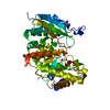
| ||||||||
|---|---|---|---|---|---|---|---|---|---|
| 1 | 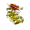
| ||||||||
| Unit cell |
| ||||||||
| Components on special symmetry positions |
|
-Components
| #1: Protein | Mass: 46876.422 Da / Num. of mol.: 1 Source method: isolated from a genetically manipulated source Details: CHAIN A HAS COVALENTLY LINKED 2 VISIBLE RESIDUES IN AN ARTEFACTUAL N-TERMINAL EXTENSION RESULTING FROM CLEAVAGE OF HIS-TAG, RESIDUES -3 TO -1 Source: (gene. exp.)  | ||||||||||
|---|---|---|---|---|---|---|---|---|---|---|---|
| #2: Chemical |   Mass: 65.409 Da / Num. of mol.: 2 / Source method: obtained synthetically / Formula: Zn Mass: 65.409 Da / Num. of mol.: 2 / Source method: obtained synthetically / Formula: Zn#3: Chemical | ChemComp-SO4 / |   Mass: 96.063 Da / Num. of mol.: 1 / Source method: obtained synthetically / Formula: SO4 Mass: 96.063 Da / Num. of mol.: 1 / Source method: obtained synthetically / Formula: SO4#4: Chemical |   Mass: 62.068 Da / Num. of mol.: 3 / Source method: obtained synthetically / Formula: C2H6O2 Mass: 62.068 Da / Num. of mol.: 3 / Source method: obtained synthetically / Formula: C2H6O2#5: Water | ChemComp-HOH / |  Mass: 18.015 Da / Num. of mol.: 239 / Source method: isolated from a natural source / Formula: H2O Mass: 18.015 Da / Num. of mol.: 239 / Source method: isolated from a natural source / Formula: H2OCompound details | CATALYSES THE CONVERSION OF 2-PHOSPHO-D-GLYCERATE TO PHOSPHOENOLPYRUVATE AND WATER. THIS PROTEIN ...CATALYSES THE CONVERSION | Sequence details | TWO ARG RESIDUES ARE MISTRANSLA | |
-Experimental details
-Experiment
| Experiment | Method: X-RAY DIFFRACTION / Number of used crystals: 1 |
|---|
- Sample preparation
| Crystal | Density Matthews: 2.38 Å3/Da / Density % sol: 47.92 % |
|---|---|
| Crystal grow | pH: 6.5 / Details: 0.01M ZNSO4,0.1 M MES PH 6.5, 25% PEGMME550 |
| Crystal grow | *PLUS Temperature: 18 ℃ / Method: vapor diffusion, hanging drop / Details: Hannaert, V., (2003) Eur.J.Biochem., 270, 3205. |
| Components of the solutions | *PLUS Conc.: 6 mg/ml / Common name: protein |
-Data collection
| Diffraction | Mean temperature: 100 K |
|---|---|
| Diffraction source | Source: ROTATING ANODE / Type: RIGAKU ULTRAX 18 / Wavelength: 1.5418 |
| Detector | Type: MARRESEARCH / Detector: IMAGE PLATE / Date: Oct 15, 2002 |
| Radiation | Protocol: SINGLE WAVELENGTH / Monochromatic (M) / Laue (L): M / Scattering type: x-ray |
| Radiation wavelength | Wavelength: 1.5418 Å / Relative weight: 1 |
| Reflection | Resolution: 2.3→38.82 Å / Num. obs: 20116 / % possible obs: 99.3 % / Redundancy: 4.7 % / Biso Wilson estimate: 32.6 Å2 / Rmerge(I) obs: 0.065 / Net I/σ(I): 14.2 |
| Reflection shell | Resolution: 2.3→2.38 Å / Redundancy: 4.5 % / Rmerge(I) obs: 0.468 / Mean I/σ(I) obs: 1.9 / % possible all: 93.4 |
| Reflection | *PLUS Lowest resolution: 38.8 Å / % possible obs: 97.2 % / Rmerge(I) obs: 0.0652 |
| Reflection shell | *PLUS % possible obs: 93.4 % |
- Processing
| Software |
| ||||||||||||||||||||||||||||||||||||||||||||||||||||||||||||||||||||||||||||||||
|---|---|---|---|---|---|---|---|---|---|---|---|---|---|---|---|---|---|---|---|---|---|---|---|---|---|---|---|---|---|---|---|---|---|---|---|---|---|---|---|---|---|---|---|---|---|---|---|---|---|---|---|---|---|---|---|---|---|---|---|---|---|---|---|---|---|---|---|---|---|---|---|---|---|---|---|---|---|---|---|---|---|
| Refinement | Method to determine structure: MOLECULAR REPLACEMENT Starting model: PDB ENTRY 1ONE Resolution: 2.3→38.82 Å / Rfactor Rfree error: 0.008 / Isotropic thermal model: RESTRAINED / Cross valid method: THROUGHOUT / σ(F): 0
| ||||||||||||||||||||||||||||||||||||||||||||||||||||||||||||||||||||||||||||||||
| Solvent computation | Solvent model: FLAT MODEL / Bsol: 41.1806 Å2 / ksol: 0.327786 e/Å3 | ||||||||||||||||||||||||||||||||||||||||||||||||||||||||||||||||||||||||||||||||
| Displacement parameters | Biso mean: 43.7 Å2
| ||||||||||||||||||||||||||||||||||||||||||||||||||||||||||||||||||||||||||||||||
| Refine analyze |
| ||||||||||||||||||||||||||||||||||||||||||||||||||||||||||||||||||||||||||||||||
| Refinement step | Cycle: LAST / Resolution: 2.3→38.82 Å
| ||||||||||||||||||||||||||||||||||||||||||||||||||||||||||||||||||||||||||||||||
| Refine LS restraints |
| ||||||||||||||||||||||||||||||||||||||||||||||||||||||||||||||||||||||||||||||||
| LS refinement shell | Resolution: 2.3→2.38 Å / Rfactor Rfree error: 0.035 / Total num. of bins used: 10
| ||||||||||||||||||||||||||||||||||||||||||||||||||||||||||||||||||||||||||||||||
| Refinement | *PLUS Num. reflection obs: 17334 / Rfactor Rwork: 0.21 | ||||||||||||||||||||||||||||||||||||||||||||||||||||||||||||||||||||||||||||||||
| Solvent computation | *PLUS | ||||||||||||||||||||||||||||||||||||||||||||||||||||||||||||||||||||||||||||||||
| Displacement parameters | *PLUS | ||||||||||||||||||||||||||||||||||||||||||||||||||||||||||||||||||||||||||||||||
| Refine LS restraints | *PLUS
| ||||||||||||||||||||||||||||||||||||||||||||||||||||||||||||||||||||||||||||||||
| LS refinement shell | *PLUS Num. reflection Rwork: 1942 |
