 Movie
Movie Controller

[English] 日本語
 Yorodumi
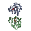
- PDB-2pt0: Structure of Selenomonas ruminantium PTP-like phytase with the ac... -
+ Open data

- Basic information
| Entry | Database: PDB / ID: 2pt0 | ||||||
|---|---|---|---|---|---|---|---|
| Title | Structure of Selenomonas ruminantium PTP-like phytase with the active site cysteine oxidized to cysteine-sulfonic acid | ||||||
 Components Components | Myo-inositol hexaphosphate phosphohydrolase | ||||||
 Keywords Keywords | HYDROLASE / PTP / protein tyrosine phosphatase / phytase / P-loop / cysteine-sulfonic acid / oxidized thiol. | ||||||
| Function / homology |  Function and homology information Function and homology information | ||||||
| Biological species |  Selenomonas ruminantium (bacteria) Selenomonas ruminantium (bacteria) | ||||||
| Method |  X-RAY DIFFRACTION / SYNCHROTRON / MOLECULAR REPLACEMENT / Resolution: 1.7 Å X-RAY DIFFRACTION / SYNCHROTRON / MOLECULAR REPLACEMENT / Resolution: 1.7 Å | ||||||
 Authors Authors | Gruninger, R.J. / Selinger, L.B. / Mosimann, S.C. | ||||||
 Citation Citation | Journal: Febs J. / Year: 2008 Title: Effect of ionic strength and oxidation on the P-loop conformation of the protein tyrosine phosphatase-like phytase, PhyAsr. Authors: Gruninger, R.J. / Brent Selinger, L. / Mosimann, S.C. | ||||||
| History |
|
- Structure visualization
| Structure viewer | Molecule: MolmilJmol/JSmol |
|---|
- Downloads & links
-Download
| PDBx/mmCIF format | 2pt0.cif.gz | 154.3 KB | Display | PDBx/mmCIF format |
|---|---|---|---|---|
| PDB format | pdb2pt0.ent.gz | 119.5 KB | Display | PDB format |
| PDBx/mmJSON format | 2pt0.json.gz | Tree view | PDBx/mmJSON format | |
| Others |  Other downloads Other downloads |
-Validation report
| Arichive directory | https://data.pdbj.org/pub/pdb/validation_reports/pt/2pt0ftp://data.pdbj.org/pub/pdb/validation_reports/pt/2pt0 | HTTPS FTP |
|---|
-Related structure data
| Related structure data |  2pszC  3d1hC  3d1oC  3d1qC  2b4pS S: Starting model for refinement C: citing same article ( |
|---|---|
| Similar structure data |


-Links
 PDBj
PDBj


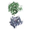
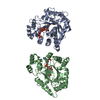
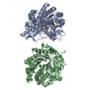
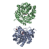
- Assembly
| Deposited unit | 
| ||||||||
|---|---|---|---|---|---|---|---|---|---|
| 1 |
| ||||||||
| Unit cell |
|
-Components
| #1: Protein | Mass: 39035.910 Da / Num. of mol.: 2 Source method: isolated from a genetically manipulated source Source: (gene. exp.) Selenomonas ruminantium (bacteria) / Gene: phyA / Plasmid: pET28b / Species (production host): Escherichia coli / Production host: #2: Chemical |   Mass: 92.094 Da / Num. of mol.: 3 / Source method: obtained synthetically / Formula: C3H8O3 Mass: 92.094 Da / Num. of mol.: 3 / Source method: obtained synthetically / Formula: C3H8O3#3: Water | ChemComp-HOH / |  Mass: 18.015 Da / Num. of mol.: 686 / Source method: isolated from a natural source / Formula: H2O Mass: 18.015 Da / Num. of mol.: 686 / Source method: isolated from a natural source / Formula: H2O |
|---|
-Experimental details
-Experiment
| Experiment | Method: X-RAY DIFFRACTION / Number of used crystals: 1 |
|---|
- Sample preparation
| Crystal | Density Matthews: 3.18 Å3/Da / Density % sol: 61.32 % |
|---|---|
| Crystal grow | Temperature: 293 K / Method: vapor diffusion, sitting drop / pH: 4.8 Details: 10% P8K, 200 mM NaCl, 50 mM Na-acetate (pH 4.8), VAPOR DIFFUSION, SITTING DROP, temperature 293K |
-Data collection
| Diffraction | Mean temperature: 100 K |
|---|---|
| Diffraction source | Source: SYNCHROTRON / Site: ALS  / Beamline: 8.3.1 / Wavelength: 1.11 Å / Beamline: 8.3.1 / Wavelength: 1.11 Å |
| Detector | Type: ADSC QUANTUM 210 / Detector: CCD / Date: Oct 20, 2006 |
| Radiation | Monochromator: double crystal / Protocol: SINGLE WAVELENGTH / Monochromatic (M) / Laue (L): M / Scattering type: x-ray |
| Radiation wavelength | Wavelength: 1.11 Å / Relative weight: 1 |
| Reflection | Resolution: 1.7→30 Å / Num. all: 95985 / Num. obs: 95985 / % possible obs: 89.8 % / Observed criterion σ(F): 1.5 / Observed criterion σ(I): 2 / Redundancy: 2.1 % / Biso Wilson estimate: 20.9 Å2 / Rmerge(I) obs: 0.038 / Rsym value: 0.041 / Net I/σ(I): 23.7 |
| Reflection shell | Resolution: 1.7→1.76 Å / Redundancy: 1.8 % / Rmerge(I) obs: 0.237 / Mean I/σ(I) obs: 3.81 / Num. unique all: 5219 / Rsym value: 0.233 / % possible all: 54 |
- Processing
| Software |
| |||||||||||||||||||||||||
|---|---|---|---|---|---|---|---|---|---|---|---|---|---|---|---|---|---|---|---|---|---|---|---|---|---|---|
| Refinement | Method to determine structure: MOLECULAR REPLACEMENT Starting model: 2B4P Resolution: 1.7→30 Å / Cross valid method: THROUGHOUT / σ(F): 2 / σ(I): 1.5 / Stereochemistry target values: Engh & Huber
| |||||||||||||||||||||||||
| Refinement step | Cycle: LAST / Resolution: 1.7→30 Å
|
