 Movie
Movie Controller

[English] 日本語
 Yorodumi
- PDB-3d1h: Structure of the PTP-Like Phytase Expressed by Selenomonas Rumina... -
+ Open data

- Basic information
| Entry | Database: PDB / ID: 3d1h | ||||||
|---|---|---|---|---|---|---|---|
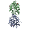
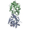
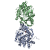
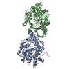
| Title | Structure of the PTP-Like Phytase Expressed by Selenomonas Ruminantium at an Ionic Strength of 500 mM | ||||||
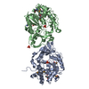
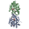
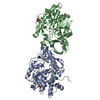
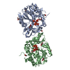
 Components Components | Myo-inositol hexaphosphate phosphohydrolase | ||||||
 Keywords Keywords | HYDROLASE / PTP / PROTEIN TYROSINE PHOSPHATASE / PHYTASE / P-LOOP | ||||||
| Function / homology |  Function and homology information Function and homology information | ||||||
| Biological species |  Selenomonas ruminantium (bacteria) Selenomonas ruminantium (bacteria) | ||||||
| Method |  X-RAY DIFFRACTION / SYNCHROTRON / MOLECULAR REPLACEMENT / Resolution: 2.1 Å X-RAY DIFFRACTION / SYNCHROTRON / MOLECULAR REPLACEMENT / Resolution: 2.1 Å | ||||||
 Authors Authors | Gruninger, R.J. / Selinger, L.B. / Mosimann, S.C. | ||||||
 Citation Citation | Journal: Febs J. / Year: 2008 Title: Effect of ionic strength and oxidation on the P-loop conformation of the protein tyrosine phosphatase-like phytase, PhyAsr. Authors: Gruninger, R.J. / Brent Selinger, L. / Mosimann, S.C. | ||||||
| History |
|
- Structure visualization
| Structure viewer | Molecule: MolmilJmol/JSmol |
|---|
- Downloads & links
-Download
| PDBx/mmCIF format | 3d1h.cif.gz | 148.3 KB | Display | PDBx/mmCIF format |
|---|---|---|---|---|
| PDB format | pdb3d1h.ent.gz | 115.5 KB | Display | PDB format |
| PDBx/mmJSON format | 3d1h.json.gz | Tree view | PDBx/mmJSON format | |
| Others |  Other downloads Other downloads |
-Validation report
| Arichive directory | https://data.pdbj.org/pub/pdb/validation_reports/d1/3d1hftp://data.pdbj.org/pub/pdb/validation_reports/d1/3d1h | HTTPS FTP |
|---|
-Related structure data
| Related structure data |  2pszC  2pt0C  3d1oC  3d1qC  2b4pS S: Starting model for refinement C: citing same article ( |
|---|---|
| Similar structure data |


-Links
 PDBj
PDBj


- Assembly
| Deposited unit | 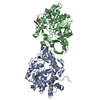
| ||||||||
|---|---|---|---|---|---|---|---|---|---|
| 1 |
| ||||||||
| Unit cell |
|
-Components
| #1: Protein | Mass: 38987.910 Da / Num. of mol.: 2 Source method: isolated from a genetically manipulated source Source: (gene. exp.) Selenomonas ruminantium (bacteria) / Gene: phyA / Plasmid: pET28b / Production host: #2: Chemical | ChemComp-GOL /   Mass: 92.094 Da / Num. of mol.: 5 / Source method: obtained synthetically / Formula: C3H8O3 Mass: 92.094 Da / Num. of mol.: 5 / Source method: obtained synthetically / Formula: C3H8O3#3: Water | ChemComp-HOH / |  Mass: 18.015 Da / Num. of mol.: 484 / Source method: isolated from a natural source / Formula: H2O Mass: 18.015 Da / Num. of mol.: 484 / Source method: isolated from a natural source / Formula: H2O |
|---|
-Experimental details
-Experiment
| Experiment | Method: X-RAY DIFFRACTION / Number of used crystals: 1 |
|---|
- Sample preparation
| Crystal | Density Matthews: 3.14 Å3/Da / Density % sol: 60.81 % |
|---|---|
| Crystal grow | Temperature: 293 K / Method: vapor diffusion, sitting drop / pH: 4.8 Details: 10% P8K, 450 MM NACL, 50 MM NA-ACETATE, pH 4.8, VAPOR DIFFUSION, SITTING DROP, temperature 293K |
-Data collection
| Diffraction | Mean temperature: 100 K |
|---|---|
| Diffraction source | Source: SYNCHROTRON / Site: ALS  / Beamline: 8.3.1 / Wavelength: 1.1158 Å / Beamline: 8.3.1 / Wavelength: 1.1158 Å |
| Detector | Type: ADSC QUANTUM 210 / Detector: CCD / Date: Oct 20, 2006 |
| Radiation | Monochromator: DOUBLE CRYSTAL / Protocol: SINGLE WAVELENGTH / Monochromatic (M) / Laue (L): M / Scattering type: x-ray |
| Radiation wavelength | Wavelength: 1.1158 Å / Relative weight: 1 |
| Reflection | Resolution: 2.1→50 Å / Num. obs: 46736 / % possible obs: 91 % / Observed criterion σ(F): 2 / Observed criterion σ(I): 2 / Redundancy: 2 % / Biso Wilson estimate: 18.8 Å2 / Rmerge(I) obs: 0.063 / Rsym value: 0.055 / Net I/σ(I): 16.6 |
| Reflection shell | Resolution: 2.1→2.18 Å / Redundancy: 1.6 % / Rmerge(I) obs: 0.207 / Mean I/σ(I) obs: 3.2 / Num. unique all: 3133 / Rsym value: 0.19 / % possible all: 56.4 |
- Processing
| Software |
| |||||||||||||||||||||||||
|---|---|---|---|---|---|---|---|---|---|---|---|---|---|---|---|---|---|---|---|---|---|---|---|---|---|---|
| Refinement | Method to determine structure: MOLECULAR REPLACEMENT Starting model: PDB entry 2b4p Resolution: 2.1→50 Å / Isotropic thermal model: isotropic / Cross valid method: THROUGHOUT / σ(F): 1.5 / σ(I): 1.5 / Stereochemistry target values: Engh & Huber
| |||||||||||||||||||||||||
| Refinement step | Cycle: LAST / Resolution: 2.1→50 Å
|
