 Movie
Movie Controller


+ Open data

- Basic information
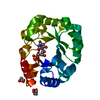
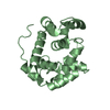
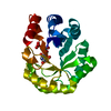
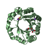
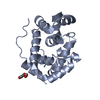
| Entry | Database: PDB / ID: 2pru | ||||||
|---|---|---|---|---|---|---|---|
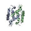
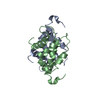
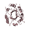
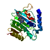
| Title | NMR Structure of Human apoS100B at 10C | ||||||
 Components Components | Protein S100-B | ||||||
 Keywords Keywords | METAL BINDING PROTEIN / S100 / Calcium Binding Protein / EF-hand / all alpha helical protein | ||||||
| Function / homology |  Function and homology information Function and homology informationnegative regulation of skeletal muscle cell differentiation / adaptive thermogenesis / sympathetic neuron projection extension / RAGE receptor binding / positive regulation of myelination / response to methylmercury / astrocyte differentiation / S100 protein binding / response to anesthetic / TRAF6 mediated NF-kB activation ...negative regulation of skeletal muscle cell differentiation / adaptive thermogenesis / sympathetic neuron projection extension / RAGE receptor binding / positive regulation of myelination / response to methylmercury / astrocyte differentiation / S100 protein binding / response to anesthetic / TRAF6 mediated NF-kB activation / Advanced glycosylation endproduct receptor signaling / regulation of neuronal synaptic plasticity / Nuclear signaling by ERBB4 / ruffle / positive regulation of neuron differentiation / axonogenesis / response to glucocorticoid / central nervous system development / TAK1-dependent IKK and NF-kappa-B activation / tau protein binding / memory / long-term synaptic potentiation / calcium-dependent protein binding / regulation of cell shape / microtubule cytoskeleton / cellular response to hypoxia / learning or memory / positive regulation of canonical NF-kappaB signal transduction / cell adhesion / cilium / ciliary basal body / positive regulation of apoptotic process / neuronal cell body / intracellular membrane-bounded organelle / positive regulation of cell population proliferation / calcium ion binding / perinuclear region of cytoplasm / protein homodimerization activity / extracellular space / extracellular region / zinc ion binding / nucleoplasm / identical protein binding / nucleus / cytosol / cytoplasm Similarity search - Function | ||||||
| Biological species |  Homo sapiens (human) Homo sapiens (human) | ||||||
| Method | SOLUTION NMR / CNS - non-crystallograqphic symmetry for the homodimer, distance geometry, simulated annealing, molecular dynamics. Final Refinement in the presence of explicit solvent. | ||||||
 Authors Authors | Malik, S. / Shaw, G.S. / Revington, M. | ||||||
 Citation Citation | Journal: Proteins / Year: 2008 Title: Analysis of the structure of human apo-S100B at low temperature indicates a unimodal conformational distribution is adopted by calcium-free S100 proteins. Authors: Malik, S. / Revington, M. / Smith, S.P. / Shaw, G.S. | ||||||
| History |
|
- Structure visualization
| Structure viewer | Molecule: MolmilJmol/JSmol |
|---|
- Downloads & links
-Download
| PDBx/mmCIF format | 2pru.cif.gz | 1.1 MB | Display | PDBx/mmCIF format |
|---|---|---|---|---|
| PDB format | pdb2pru.ent.gz | 939.6 KB | Display | PDB format |
| PDBx/mmJSON format | 2pru.json.gz | Tree view | PDBx/mmJSON format | |
| Others |  Other downloads Other downloads |
-Validation report
| Summary document | 2pru_validation.pdf.gz | 355 KB | Display | wwPDB validaton report |
|---|---|---|---|---|
| Full document | 2pru_full_validation.pdf.gz | 603.1 KB | Display | |
| Data in XML | 2pru_validation.xml.gz | 60.9 KB | Display | |
| Data in CIF | 2pru_validation.cif.gz | 92.7 KB | Display | |
| Arichive directory | https://data.pdbj.org/pub/pdb/validation_reports/pr/2pruftp://data.pdbj.org/pub/pdb/validation_reports/pr/2pru | HTTPS FTP |
-Related structure data
| Related structure data | |
|---|---|
| Similar structure data |


-Links
 PDBj
PDBj


- Assembly
| Deposited unit | 
| |||||||||
|---|---|---|---|---|---|---|---|---|---|---|
| 1 |
| |||||||||
| NMR ensembles |
|
-Components
| #1: Protein | Mass: 10595.841 Da / Num. of mol.: 2 Source method: isolated from a genetically manipulated source Source: (gene. exp.) Homo sapiens (human) / Gene: S100B / Plasmid: pSS2 from pSD80 / Production host:  |
|---|
-Experimental details
-Experiment
| Experiment | Method: SOLUTION NMR | ||||||||||||||||
|---|---|---|---|---|---|---|---|---|---|---|---|---|---|---|---|---|---|
| NMR experiment |
| ||||||||||||||||
| NMR details | Text: The structure was determined using triple-resonance NMR spectroscopy. |
 HSQC
HSQC- Sample preparation
| Details |
| |||||||||
|---|---|---|---|---|---|---|---|---|---|---|
| Sample conditions | Ionic strength: 50 mM / pH: 7.2 / Pressure: ambient / Temperature: 283 K |
-NMR measurement
| NMR spectrometer |
|
|---|
- Processing
| NMR software |
| ||||||||||||||||||||||||
|---|---|---|---|---|---|---|---|---|---|---|---|---|---|---|---|---|---|---|---|---|---|---|---|---|---|
| Refinement | Method: CNS - non-crystallograqphic symmetry for the homodimer, distance geometry, simulated annealing, molecular dynamics. Final Refinement in the presence of explicit solvent. Software ordinal: 1 / Details: 2504 NOEs, 124 Hbond distance restraints | ||||||||||||||||||||||||
| NMR representative | Selection criteria: closest to the average | ||||||||||||||||||||||||
| NMR ensemble | Conformer selection criteria: structures with the lowest energy Conformers calculated total number: 100 / Conformers submitted total number: 20 |
