 Movie
Movie Controller

[English] 日本語
 Yorodumi
- PDB-6ghy: Structure of Lytic Transglycosylase MltE inactive mutant E64Q fro... -
+ Open data

- Basic information
| Entry | Database: PDB / ID: 6ghy | ||||||
|---|---|---|---|---|---|---|---|
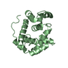
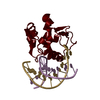
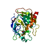
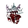
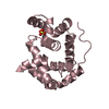
| Title | Structure of Lytic Transglycosylase MltE inactive mutant E64Q from E.coli | ||||||
 Components Components | Endo-type membrane-bound lytic murein transglycosylase A | ||||||
 Keywords Keywords | LYASE / Lytic Transglycosylase | ||||||
| Function / homology |  Function and homology information Function and homology information: / lytic endotransglycosylase activity / peptidoglycan lytic transglycosylase activity / peptidoglycan metabolic process / cell outer membrane / cell wall organization / cell wall macromolecule catabolic process / cell division Similarity search - Function | ||||||
| Biological species |  | ||||||
| Method |  X-RAY DIFFRACTION / SYNCHROTRON / MOLECULAR REPLACEMENT / Resolution: 2.12 Å X-RAY DIFFRACTION / SYNCHROTRON / MOLECULAR REPLACEMENT / Resolution: 2.12 Å | ||||||
 Authors Authors | Batuecas, M.T. / Hermoso, J.A. | ||||||
| Funding support |  Spain, 1items Spain, 1items
| ||||||
 Citation Citation | Journal: Biochemistry / Year: 2018 Title: A Structural Dissection of the Active Site of the Lytic Transglycosylase MltE from Escherichia coli. Authors: Dik, D.A. / Batuecas, M.T. / Lee, M. / Mahasenan, K.V. / Marous, D.R. / Lastochkin, E. / Fisher, J.F. / Hermoso, J.A. / Mobashery, S. | ||||||
| History |
|
- Structure visualization
| Structure viewer | Molecule: MolmilJmol/JSmol |
|---|
- Downloads & links
-Download
| PDBx/mmCIF format | 6ghy.cif.gz | 92.6 KB | Display | PDBx/mmCIF format |
|---|---|---|---|---|
| PDB format | pdb6ghy.ent.gz | 70.7 KB | Display | PDB format |
| PDBx/mmJSON format | 6ghy.json.gz | Tree view | PDBx/mmJSON format | |
| Others |  Other downloads Other downloads |
-Validation report
| Arichive directory | https://data.pdbj.org/pub/pdb/validation_reports/gh/6ghyftp://data.pdbj.org/pub/pdb/validation_reports/gh/6ghy | HTTPS FTP |
|---|
-Related structure data
| Related structure data |  6ghzC  6gi3C  6gi4C  2y8pS S: Starting model for refinement C: citing same article ( |
|---|---|
| Similar structure data |


-Links
 PDBj
PDBj


- Assembly
| Deposited unit | 
| |||||||||
|---|---|---|---|---|---|---|---|---|---|---|
| 1 | 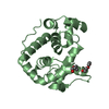
| |||||||||
| 2 | 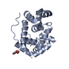
| |||||||||
| Unit cell |
| |||||||||
| Components on special symmetry positions |
|
-Components
| #1: Protein | Mass: 21413.326 Da / Num. of mol.: 2 Source method: isolated from a genetically manipulated source Source: (gene. exp.) Strain: K12 / Gene: emtA, mltE, sltZ, ycgP, b1193, JW5821 / Production host: References: UniProt: P0C960, Lyases; Carbon-oxygen lyases; Acting on polysaccharides #2: Chemical |   Mass: 106.120 Da / Num. of mol.: 3 / Source method: obtained synthetically / Formula: C4H10O3 Mass: 106.120 Da / Num. of mol.: 3 / Source method: obtained synthetically / Formula: C4H10O3#3: Water | ChemComp-HOH / |  Mass: 18.015 Da / Num. of mol.: 295 / Source method: isolated from a natural source / Formula: H2O Mass: 18.015 Da / Num. of mol.: 295 / Source method: isolated from a natural source / Formula: H2O |
|---|
-Experimental details
-Experiment
| Experiment | Method: X-RAY DIFFRACTION / Number of used crystals: 1 |
|---|
- Sample preparation
| Crystal | Density Matthews: 2.24 Å3/Da / Density % sol: 45.02 % |
|---|---|
| Crystal grow | Temperature: 291 K / Method: batch mode / pH: 8.4 Details: 26% Polyethylene Glycol 4000, 0.2 M magnesium chloride and 0.1 M TRIS HCl (pH 8.4) |
-Data collection
| Diffraction | Mean temperature: 100 K |
|---|---|
| Diffraction source | Source: SYNCHROTRON / Site: ALBA / Beamline: XALOC / Wavelength: 0.97242 Å |
| Detector | Type: DECTRIS PILATUS 6M / Detector: PIXEL / Date: Feb 12, 2016 |
| Radiation | Protocol: SINGLE WAVELENGTH / Monochromatic (M) / Laue (L): M / Scattering type: x-ray |
| Radiation wavelength | Wavelength: 0.97242 Å / Relative weight: 1 |
| Reflection | Resolution: 2.12→39.55 Å / Num. obs: 22063 / % possible obs: 98.06 % / Redundancy: 4.2 % / Rpim(I) all: 0.047 / Net I/σ(I): 11 |
| Reflection shell | Resolution: 2.12→2.19 Å |
- Processing
| Software |
| ||||||||||||||||||||
|---|---|---|---|---|---|---|---|---|---|---|---|---|---|---|---|---|---|---|---|---|---|
| Refinement | Method to determine structure: MOLECULAR REPLACEMENT Starting model: 2Y8P Resolution: 2.12→39.55 Å / Cor.coef. Fo:Fc: 0.95 / Cor.coef. Fo:Fc free: 0.929 / SU B: 0.003 / SU ML: 0 / Cross valid method: THROUGHOUT / ESU R: 0.173 / ESU R Free: 0.209 / Details: HYDROGENS HAVE BEEN ADDED IN THE RIDING POSITIONS
| ||||||||||||||||||||
| Solvent computation | Ion probe radii: 0.8 Å / Shrinkage radii: 0.8 Å / VDW probe radii: 1.2 Å | ||||||||||||||||||||
| Displacement parameters | Biso mean: 35.334 Å2
| ||||||||||||||||||||
| Refinement step | Cycle: 1 / Resolution: 2.12→39.55 Å
| ||||||||||||||||||||
| LS refinement shell | Resolution: 2.12→2.175 Å / Total num. of bins used: 20
|
