- PDB-2prr: Crystal structure of alkylhydroperoxidase AhpD core: uncharacteri... -
+
Open data
ID or keywords:
Loading...
-
Basic information
Entry
Database: PDB / ID: 2prr
Title
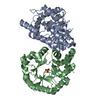
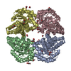
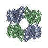
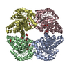
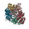
Crystal structure of alkylhydroperoxidase AhpD core: uncharacterized peroxidase-related protein (YP_296737.1) from Ralstonia eutropha JMP134 at 2.15 A resolution
Components
Alkylhydroperoxidase AhpD core: uncharacterized peroxidase-related protein
Keywords
OXIDOREDUCTASE / YP_296737.1 / Carboxymuconolactone decarboxylase family / Alkylhydroperoxidase AhpD core: uncharacterized peroxidase-related / Structural Genomics / Joint Center for Structural Genomics / JCSG / Protein Structure Initiative / PSI-2
BIOMOLECULE: 1,2 THIS ENTRY CONTAINS THE CRYSTALLOGRAPHIC ASYMMETRIC UNIT WHICH CONSISTS OF 12 ... BIOMOLECULE: 1,2 THIS ENTRY CONTAINS THE CRYSTALLOGRAPHIC ASYMMETRIC UNIT WHICH CONSISTS OF 12 CHAIN(S). SEE REMARK 350 FOR INFORMATION ON GENERATING THE BIOLOGICAL MOLECULE(S).
Remark 999
SEQUENCE THE CONSTRUCT WAS EXPRESSED WITH A PURIFICATION TAG MGSDKIHHHHHHENLYFQG. THE TAG WAS ... SEQUENCE THE CONSTRUCT WAS EXPRESSED WITH A PURIFICATION TAG MGSDKIHHHHHHENLYFQG. THE TAG WAS REMOVED WITH TEV PROTEASE LEAVING ONLY A GLYCINE (0) FOLLOWED BY THE TARGET SEQUENCE.
Type: MARMOSAIC 325 mm CCD / Detector: CCD / Date: Dec 4, 2006 / Details: Flat mirror (vertical focusing)
Radiation
Monochromator: Single crystal Si(111) bent (horizontal focusing) Protocol: MAD / Monochromatic (M) / Laue (L): M / Scattering type: x-ray
Radiation wavelength
ID
Wavelength (Å)
Relative weight
1
0.91837
1
2
0.97932
1
3
0.97905
1
Reflection
Resolution: 1.88→48.912 Å / Num. obs: 211872 / % possible obs: 87.2 % / Biso Wilson estimate: 30.222 Å2 / Rmerge(I) obs: 0.079 / Net I/σ(I): 6.23
Reflection shell
Diffraction-ID: 1
Resolution (Å)
Highest resolution (Å)
Rmerge(I) obs
Mean I/σ(I) obs
Num. measured obs
Num. unique all
% possible all
1.88-1.95
0.419
2
35774
18037
77.4
1.95-2.03
0.378
2.2
37831
19032
83.1
2.03-2.12
0.274
2.8
36081
18150
83.4
2.12-2.23
0.235
3.2
38632
19422
87.9
2.23-2.37
0.174
4.2
38384
19293
86.1
2.37-2.55
0.137
5
39147
19674
88.9
2.55-2.81
0.109
6.2
40063
20136
88.7
2.81-3.21
0.076
8.2
39288
19738
89.1
3.21
0.047
11.9
41650
20939
93.1
-
Phasing
Phasing
Method: MAD
-
Processing
Software
Name
Version
Classification
NB
MolProbity
3beta29
modelbuilding
SHELX
phasing
REFMAC
5.2.0005
refinement
XSCALE
datascaling
PDB_EXTRACT
2
dataextraction
MAR345
CCD
datacollection
XDS
datareduction
SHELXD
phasing
autoSHARP
phasing
Refinement
Method to determine structure: MAD / Resolution: 2.15→48.912 Å / Cor.coef. Fo:Fc: 0.949 / Cor.coef. Fo:Fc free: 0.917 / SU B: 8.887 / SU ML: 0.21 / Cross valid method: THROUGHOUT / σ(F): 0 / ESU R: 0.245 / ESU R Free: 0.208 Stereochemistry target values: MAXIMUM LIKELIHOOD WITH PHASES Details: 1. HYDROGENS HAVE BEEN ADDED IN THE RIDING POSITIONS. 2. ATOM RECORD CONTAINS RESIDUAL B FACTORS ONLY. 3. A MET-INHIBITION PROTOCOL WAS USED FOR SELENOMETHIONINE INCORPORATION DURING PROTEIN ...Details: 1. HYDROGENS HAVE BEEN ADDED IN THE RIDING POSITIONS. 2. ATOM RECORD CONTAINS RESIDUAL B FACTORS ONLY. 3. A MET-INHIBITION PROTOCOL WAS USED FOR SELENOMETHIONINE INCORPORATION DURING PROTEIN EXPRESSION. THE OCCUPANCY OF THE SE ATOMS IN THE MSE RESIDUES WAS REDUCED TO 0.75 TO ACCOUNT FOR THE REDUCED SCATTERING POWER DUE TO PARTIAL S-MET INCORPORATION. 4. POLYETHYLENE GLYCOL MOLECULES FROM THE CRYSTALLIZATION BUFFER WERE MODELED INTO THE STRUCTURE.
Rfactor
Num. reflection
% reflection
Selection details
Rfree
0.262
7726
5.1 %
RANDOM
Rwork
0.211
-
-
-
all
0.213
-
-
-
obs
0.213
152688
94.68 %
-
Solvent computation
Ion probe radii: 0.8 Å / Shrinkage radii: 0.8 Å / VDW probe radii: 1.2 Å / Solvent model: BABINET MODEL WITH MASK
 Movie
Movie Controller

 Yorodumi
 Open data
 Basic information
 Components
 Keywords
 Function and homology information
 Ralstonia eutropha (bacteria)
 X-RAY DIFFRACTION /
  Authors
 Citation
 Structure visualization
 Downloads & links
 Other downloads
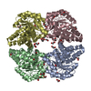


 PDBj
PDBj Assembly


 Mass: 106.120 Da / Num. of mol.: 25 / Source method: obtained synthetically / Formula: C4H10O3
 Mass: 150.173 Da / Num. of mol.: 7 / Source method: obtained synthetically / Formula: C6H14O4
 Mass: 18.015 Da / Num. of mol.: 1338 / Source method: isolated from a natural source / Formula: H2O
 Sample preparation
 / Beamline: BL11-1 / Wavelength: 0.91837, 0.97932, 0.97905
 Processing