 Movie
Movie Controller


+ Open data

- Basic information
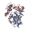
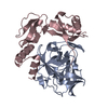
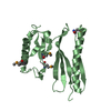
| Entry | Database: PDB / ID: 2pro | ||||||
|---|---|---|---|---|---|---|---|
| Title | PRO REGION OF ALPHA-LYTIC PROTEASE | ||||||
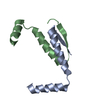
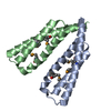
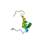
 Components Components | ALPHA-LYTIC PROTEASE | ||||||
 Keywords Keywords | PRO REGION / FOLDASE / PROTEIN FOLDING / SERINE PROTEASE | ||||||
| Function / homology |  Function and homology information Function and homology informationalpha-lytic endopeptidase / serine-type endopeptidase activity / proteolysis / extracellular region Similarity search - Function | ||||||
| Biological species |  Lysobacter enzymogenes (bacteria) Lysobacter enzymogenes (bacteria) | ||||||
| Method |  X-RAY DIFFRACTION / SYNCHROTRON / MAD / Resolution: 3 Å X-RAY DIFFRACTION / SYNCHROTRON / MAD / Resolution: 3 Å | ||||||
 Authors Authors | Sauter, N.K. / Mau, T. / Rader, S.D. / Agard, D.A. | ||||||
 Citation Citation | Journal: Nat.Struct.Biol. / Year: 1998 Title: Structure of alpha-lytic protease complexed with its pro region. Authors: Sauter, N.K. / Mau, T. / Rader, S.D. / Agard, D.A. | ||||||
| History |
|
- Structure visualization
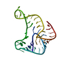
| Structure viewer | Molecule: MolmilJmol/JSmol |
|---|
- Downloads & links
-Download
| PDBx/mmCIF format | 2pro.cif.gz | 87.7 KB | Display | PDBx/mmCIF format |
|---|---|---|---|---|
| PDB format | pdb2pro.ent.gz | 68 KB | Display | PDB format |
| PDBx/mmJSON format | 2pro.json.gz | Tree view | PDBx/mmJSON format | |
| Others |  Other downloads Other downloads |
-Validation report
| Arichive directory | https://data.pdbj.org/pub/pdb/validation_reports/pr/2proftp://data.pdbj.org/pub/pdb/validation_reports/pr/2pro | HTTPS FTP |
|---|
-Related structure data


-Links
 PDBj
PDBj- Assembly
| Deposited unit | 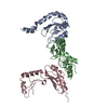
| ||||||||||||
|---|---|---|---|---|---|---|---|---|---|---|---|---|---|
| 1 | 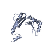
| ||||||||||||
| 2 | 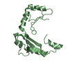
| ||||||||||||
| 3 | 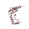
| ||||||||||||
| 4 | 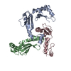
| ||||||||||||
| Unit cell |
| ||||||||||||
| Noncrystallographic symmetry (NCS) | NCS oper:
|
-Components
| #1: Protein | Mass: 17835.881 Da / Num. of mol.: 3 / Fragment: PRO REGION Source method: isolated from a genetically manipulated source Source: (gene. exp.) Lysobacter enzymogenes (bacteria) / Description: T7 EXPRESSION SYSTEM / Cell line: B834 / Plasmid: PT7PRO / Cellular location (production host): INCLUSION BODIES / Production host: Sequence details | THE FIVE METHIONINE RESIDUES IN THIS ENTRY ARE ACTUALLY A MIXTURE OF ABOUT 80% SELENO-L-METHIONINE ...THE FIVE METHIONINE | |
|---|
-Experimental details
-Experiment
| Experiment | Method: X-RAY DIFFRACTION / Number of used crystals: 1 |
|---|
- Sample preparation
| Crystal | Density Matthews: 2.94 Å3/Da / Density % sol: 58 % / Description: DATA WERE COLLECTED WITH INVERSE BEAM GEOMETRY | ||||||||||||||||||||||||||||||||||||||||||||||||||||||||||||||||||||||||||||||
|---|---|---|---|---|---|---|---|---|---|---|---|---|---|---|---|---|---|---|---|---|---|---|---|---|---|---|---|---|---|---|---|---|---|---|---|---|---|---|---|---|---|---|---|---|---|---|---|---|---|---|---|---|---|---|---|---|---|---|---|---|---|---|---|---|---|---|---|---|---|---|---|---|---|---|---|---|---|---|---|
| Crystal grow | pH: 8 / Details: pH 8.0 | ||||||||||||||||||||||||||||||||||||||||||||||||||||||||||||||||||||||||||||||
| Crystal | *PLUS | ||||||||||||||||||||||||||||||||||||||||||||||||||||||||||||||||||||||||||||||
| Crystal grow | *PLUS Temperature: 7 ℃ / Method: vapor diffusion / Details: used to seeding | ||||||||||||||||||||||||||||||||||||||||||||||||||||||||||||||||||||||||||||||
| Components of the solutions | *PLUS
|
-Data collection
| Diffraction | Mean temperature: 100 K | ||||||||||||
|---|---|---|---|---|---|---|---|---|---|---|---|---|---|
| Diffraction source | Source: SYNCHROTRON / Site: NSLS  / Beamline: X4A / Wavelength: 0.9690, 0.97915, 0.97938 / Beamline: X4A / Wavelength: 0.9690, 0.97915, 0.97938 | ||||||||||||
| Detector | Type: FUJI / Detector: IMAGE PLATE / Date: Mar 23, 1996 | ||||||||||||
| Radiation | Protocol: MAD / Monochromatic (M) / Laue (L): M / Scattering type: x-ray | ||||||||||||
| Radiation wavelength |
| ||||||||||||
| Reflection | Resolution: 3→16 Å / Num. obs: 22778 / % possible obs: 93.5 % / Redundancy: 1.6 % / Rmerge(I) obs: 0.052 / Net I/σ(I): 13 | ||||||||||||
| Reflection shell | Resolution: 3→3.14 Å / Redundancy: 1.6 % / Rmerge(I) obs: 0.222 / Mean I/σ(I) obs: 3.4 / % possible all: 94.9 | ||||||||||||
| Reflection | *PLUS Num. measured all: 39904 | ||||||||||||
| Reflection shell | *PLUS % possible obs: 94.9 % |
- Processing
| Software |
| ||||||||||||||||||||||||||||||||||||||||||||||||||||||||||||
|---|---|---|---|---|---|---|---|---|---|---|---|---|---|---|---|---|---|---|---|---|---|---|---|---|---|---|---|---|---|---|---|---|---|---|---|---|---|---|---|---|---|---|---|---|---|---|---|---|---|---|---|---|---|---|---|---|---|---|---|---|---|
| Refinement | Method to determine structure: MAD / Resolution: 3→6 Å / Cross valid method: THROUGHOUT / σ(F): 0
| ||||||||||||||||||||||||||||||||||||||||||||||||||||||||||||
| Refinement step | Cycle: LAST / Resolution: 3→6 Å
| ||||||||||||||||||||||||||||||||||||||||||||||||||||||||||||
| Refine LS restraints |
| ||||||||||||||||||||||||||||||||||||||||||||||||||||||||||||
| LS refinement shell | Resolution: 3→3.12 Å / Rfactor Rfree: 0.489 / Rfactor Rwork: 0.314 / Total num. of bins used: 8 | ||||||||||||||||||||||||||||||||||||||||||||||||||||||||||||
| Xplor file |
|
