 Movie
Movie Controller

[English] 日本語
 Yorodumi
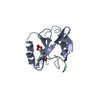
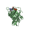
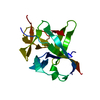
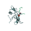
- PDB-2pld: NUCLEAR MAGNETIC RESONANCE STRUCTURE OF AN SH2 DOMAIN OF PHOSPHOL... -
+ Open data

- Basic information
| Entry | Database: PDB / ID: 2pld | ||||||
|---|---|---|---|---|---|---|---|
| Title | NUCLEAR MAGNETIC RESONANCE STRUCTURE OF AN SH2 DOMAIN OF PHOSPHOLIPASE C-GAMMA1 COMPLEXED WITH A HIGH AFFINITY BINDING PEPTIDE | ||||||
 Components Components |
| ||||||
 Keywords Keywords | PHOSPHORIC DIESTER HYDROLASE | ||||||
| Function / homology |  Function and homology information Function and homology informationplatelet-derived growth factor receptor activity / cell migration involved in coronary angiogenesis / metanephric glomerular mesangial cell proliferation involved in metanephros development / platelet activating factor receptor activity / platelet-derived growth factor beta-receptor activity / cell migration involved in vasculogenesis / positive regulation of metanephric mesenchymal cell migration by platelet-derived growth factor receptor-beta signaling pathway / : / metanephric glomerular capillary formation / smooth muscle cell chemotaxis ...platelet-derived growth factor receptor activity / cell migration involved in coronary angiogenesis / metanephric glomerular mesangial cell proliferation involved in metanephros development / platelet activating factor receptor activity / platelet-derived growth factor beta-receptor activity / cell migration involved in vasculogenesis / positive regulation of metanephric mesenchymal cell migration by platelet-derived growth factor receptor-beta signaling pathway / : / metanephric glomerular capillary formation / smooth muscle cell chemotaxis / phosphatidylinositol phospholipase C activity / smooth muscle adaptation / aorta morphogenesis / platelet-derived growth factor binding / positive regulation of cell proliferation by VEGF-activated platelet derived growth factor receptor signaling pathway / phosphoinositide phospholipase C / vascular endothelial growth factor binding / retina vasculature development in camera-type eye / phosphatidylinositol metabolic process / phosphatidylinositol-4,5-bisphosphate phospholipase C activity / cardiac myofibril assembly / Signaling by PDGF / COP9 signalosome / positive regulation of chemotaxis / phospholipase C activator activity / platelet-derived growth factor receptor binding / phospholipid catabolic process / positive regulation of smooth muscle cell migration / positive regulation of DNA biosynthetic process / platelet-derived growth factor receptor-beta signaling pathway / positive regulation of calcium ion import / platelet-derived growth factor receptor signaling pathway / positive regulation of MAP kinase activity / phosphatidylinositol-mediated signaling / positive regulation of epithelial cell migration / cellular response to vascular endothelial growth factor stimulus / ruffle / peptidyl-tyrosine phosphorylation / release of sequestered calcium ion into cytosol / positive regulation of mitotic nuclear division / lysosomal lumen / positive regulation of calcium-mediated signaling / Downstream signal transduction / positive regulation of smooth muscle cell proliferation / guanyl-nucleotide exchange factor activity / cell surface receptor protein tyrosine kinase signaling pathway / cellular response to epidermal growth factor stimulus / GTPase activator activity / regulation of actin cytoskeleton organization / cell chemotaxis / receptor protein-tyrosine kinase / positive regulation of reactive oxygen species metabolic process / epidermal growth factor receptor signaling pathway / ruffle membrane / Constitutive Signaling by Aberrant PI3K in Cancer / protein autophosphorylation / PIP3 activates AKT signaling / lamellipodium / PI5P, PP2A and IER3 Regulate PI3K/AKT Signaling / RAF/MAP kinase cascade / protein tyrosine kinase activity / angiogenesis / cytoplasmic vesicle / in utero embryonic development / protein kinase activity / positive regulation of ERK1 and ERK2 cascade / positive regulation of phosphatidylinositol 3-kinase/protein kinase B signal transduction / signaling receptor complex / apical plasma membrane / positive regulation of cell migration / signaling receptor binding / focal adhesion / calcium ion binding / positive regulation of cell population proliferation / protein kinase binding / enzyme binding / Golgi apparatus / signal transduction / ATP binding / membrane / nucleus / plasma membrane / cytoplasm Similarity search - Function | ||||||
| Biological species |  | ||||||
| Method | SOLUTION NMR | ||||||
 Authors Authors | Pascal, S.M. / Singer, A.U. / Gish, G. / Yamazaki, T. / Shoelson, S.E. / Pawson, T. / Kay, L.E. / Forman-Kay, J.D. | ||||||
 Citation Citation | Journal: Cell(Cambridge,Mass.) / Year: 1994 Title: Nuclear magnetic resonance structure of an SH2 domain of phospholipase C-gamma 1 complexed with a high affinity binding peptide. Authors: Pascal, S.M. / Singer, A.U. / Gish, G. / Yamazaki, T. / Shoelson, S.E. / Pawson, T. / Kay, L.E. / Forman-Kay, J.D. | ||||||
| History |
|
- Structure visualization
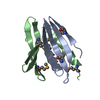
| Structure viewer | Molecule: MolmilJmol/JSmol |
|---|
- Downloads & links
-Download
| PDBx/mmCIF format | 2pld.cif.gz | 55.5 KB | Display | PDBx/mmCIF format |
|---|---|---|---|---|
| PDB format | pdb2pld.ent.gz | 39.1 KB | Display | PDB format |
| PDBx/mmJSON format | 2pld.json.gz | Tree view | PDBx/mmJSON format | |
| Others |  Other downloads Other downloads |
-Validation report
| Arichive directory | https://data.pdbj.org/pub/pdb/validation_reports/pl/2pldftp://data.pdbj.org/pub/pdb/validation_reports/pl/2pld | HTTPS FTP |
|---|
-Related structure data


-Links
 PDBj
PDBj


- Assembly
| Deposited unit | 
| |||||||||
|---|---|---|---|---|---|---|---|---|---|---|
| 1 |
| |||||||||
| Atom site foot note | 1: RESIDUES A 1 - A 10, A 99 - A 105, B 1 - B 2, AND B 11 - B 12 ARE DISORDERED IN SOLUTION; THEREFORE, COORDINATES DISPLAY LARGE RMSD VALUES FOR THESE ATOMS. | |||||||||
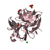
| NMR ensembles |
|
-Components
| #1: Protein | Mass: 12275.924 Da / Num. of mol.: 1 Source method: isolated from a genetically manipulated source Source: (gene. exp.) References: UniProt: P08487, phosphoinositide phospholipase C |
|---|---|
| #2: Protein/peptide | Mass: 1480.532 Da / Num. of mol.: 1 Source method: isolated from a genetically manipulated source Source: (gene. exp.) |
| Has protein modification | Y |
-Experimental details
-Experiment
| Experiment | Method: SOLUTION NMR |
|---|
- Sample preparation
| Crystal grow | *PLUS Method: other / Details: NMR |
|---|
- Processing
| Software |
| ||||||||
|---|---|---|---|---|---|---|---|---|---|
| NMR software | Name:  X-PLOR / Developer: BRUNGER / Classification: refinement X-PLOR / Developer: BRUNGER / Classification: refinement | ||||||||
| NMR ensemble | Conformers submitted total number: 1 |
