 Movie
Movie Controller


+ Open data

- Basic information
| Entry | Database: PDB / ID: 2pfb | ||||||
|---|---|---|---|---|---|---|---|
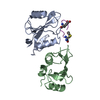
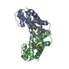
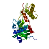
| Title | Structure of oxidized OhrR from Xanthamonas campestris | ||||||
 Components Components | Transcriptional regulator OhrR | ||||||
 Keywords Keywords | TRANSCRIPTION REGULATOR / transcriptional regulator / MarR family | ||||||
| Function / homology |  Function and homology information Function and homology informationresponse to stress / DNA-binding transcription factor activity / DNA-templated transcription / DNA binding / cytoplasm Similarity search - Function | ||||||
| Biological species |  Xanthomonas campestris (bacteria) Xanthomonas campestris (bacteria) | ||||||
| Method |  X-RAY DIFFRACTION / MOLECULAR REPLACEMENT / Resolution: 1.93 Å X-RAY DIFFRACTION / MOLECULAR REPLACEMENT / Resolution: 1.93 Å | ||||||
 Authors Authors | Brennan, R.G. / Newberry, K.J. | ||||||
 Citation Citation | Journal: Mol.Cell / Year: 2007 Title: Structural Mechanism of Organic Hydroperoxide Induction of the Transcription Regulator OhrR. Authors: Newberry, K.J. / Fuangthong, M. / Panmanee, W. / Mongkolsuk, S. / Brennan, R.G. #1: Journal: J.Bacteriol. / Year: 2006Title: Novel organic hydroperoxide-sensing and responding mechanisms for OhrR, a major bacterial sensor and regulator of organic hydroperoxide stress Authors: Panmanee, W. / Vattanaviboon, P. / Poole, L.B. / Mongkolsuk, S. | ||||||
| History |
|
- Structure visualization
| Structure viewer | Molecule: MolmilJmol/JSmol |
|---|
- Downloads & links
-Download
| PDBx/mmCIF format | 2pfb.cif.gz | 63.7 KB | Display | PDBx/mmCIF format |
|---|---|---|---|---|
| PDB format | pdb2pfb.ent.gz | 46.3 KB | Display | PDB format |
| PDBx/mmJSON format | 2pfb.json.gz | Tree view | PDBx/mmJSON format | |
| Others |  Other downloads Other downloads |
-Validation report
| Arichive directory | https://data.pdbj.org/pub/pdb/validation_reports/pf/2pfbftp://data.pdbj.org/pub/pdb/validation_reports/pf/2pfb | HTTPS FTP |
|---|
-Related structure data
| Related structure data |  2pexSC S: Starting model for refinement C: citing same article ( |
|---|---|
| Similar structure data |


-Links
 PDBj
PDBj- Assembly
| Deposited unit | 
| ||||||||
|---|---|---|---|---|---|---|---|---|---|
| 1 |
| ||||||||
| Unit cell |
| ||||||||
| Details | the biological assembly is a dimer |
-Components
| #1: Protein | Mass: 16938.436 Da / Num. of mol.: 2 / Mutation: C131S Source method: isolated from a genetically manipulated source Source: (gene. exp.) Xanthomonas campestris (bacteria) / Gene: ohrR / Plasmid: PET11a / Species (production host): Escherichia coli / Production host: #2: Water | ChemComp-HOH / |  Mass: 18.015 Da / Num. of mol.: 84 / Source method: isolated from a natural source / Formula: H2O Mass: 18.015 Da / Num. of mol.: 84 / Source method: isolated from a natural source / Formula: H2OHas protein modification | Y | |
|---|
-Experimental details
-Experiment
| Experiment | Method: X-RAY DIFFRACTION / Number of used crystals: 1 |
|---|
- Sample preparation
| Crystal | Density Matthews: 1.97 Å3/Da / Density % sol: 37.57 % |
|---|---|
| Crystal grow | Temperature: 298 K / Method: vapor diffusion, hanging drop / pH: 8.5 Details: 30% PEG 4000, 300mM magnesium chloride, 100 mM Tris, pH8.5, VAPOR DIFFUSION, HANGING DROP, temperature 298K |
-Data collection
| Diffraction | Mean temperature: 93 K |
|---|---|
| Diffraction source | Source: ROTATING ANODE / Type: RIGAKU / Wavelength: 1.5418 |
| Detector | Type: RIGAKU RAXIS HTC / Detector: IMAGE PLATE / Date: Oct 13, 2006 / Details: mirrors |
| Radiation | Monochromator: mirrors / Protocol: SINGLE WAVELENGTH / Monochromatic (M) / Laue (L): M / Scattering type: x-ray |
| Radiation wavelength | Wavelength: 1.5418 Å / Relative weight: 1 |
| Reflection | Resolution: 1.93→33.76 Å / Num. all: 20491 / Num. obs: 20477 / % possible obs: 99.9 % / Observed criterion σ(I): 3 / Redundancy: 6.5 % / Biso Wilson estimate: 40.3 Å2 / Rmerge(I) obs: 0.062 / Rsym value: 0.062 / Net I/σ(I): 14 |
| Reflection shell | Resolution: 1.93→2 Å / Redundancy: 6.13 % / Rmerge(I) obs: 0.28 / Mean I/σ(I) obs: 4.8 / Num. unique all: 2012 / Rsym value: 0.28 / % possible all: 100 |
-Phasing
| Phasing MR |
|
|---|
- Processing
| Software |
| ||||||||||||||||||||||||||||||||||||||||||||||||||||||||||||||||||||||||||||||||||||||||||||||||||||||||||||||||||
|---|---|---|---|---|---|---|---|---|---|---|---|---|---|---|---|---|---|---|---|---|---|---|---|---|---|---|---|---|---|---|---|---|---|---|---|---|---|---|---|---|---|---|---|---|---|---|---|---|---|---|---|---|---|---|---|---|---|---|---|---|---|---|---|---|---|---|---|---|---|---|---|---|---|---|---|---|---|---|---|---|---|---|---|---|---|---|---|---|---|---|---|---|---|---|---|---|---|---|---|---|---|---|---|---|---|---|---|---|---|---|---|---|---|---|---|
| Refinement | Method to determine structure: MOLECULAR REPLACEMENT Starting model: residues 18 to 107 from pdb entry 2PEX Resolution: 1.93→33.76 Å / Rfactor Rfree error: 0.008 / FOM work R set: 0.84 / Data cutoff high absF: 1872286.375 / Data cutoff low absF: 0 / Isotropic thermal model: RESTRAINED / Cross valid method: THROUGHOUT / σ(F): 0 / Stereochemistry target values: Engh & Huber / Details: BULK SOLVENT MODEL USED
| ||||||||||||||||||||||||||||||||||||||||||||||||||||||||||||||||||||||||||||||||||||||||||||||||||||||||||||||||||
| Solvent computation | Solvent model: FLAT MODEL / Bsol: 53.354 Å2 / ksol: 0.4 e/Å3 | ||||||||||||||||||||||||||||||||||||||||||||||||||||||||||||||||||||||||||||||||||||||||||||||||||||||||||||||||||
| Displacement parameters | Biso mean: 32.6 Å2
| ||||||||||||||||||||||||||||||||||||||||||||||||||||||||||||||||||||||||||||||||||||||||||||||||||||||||||||||||||
| Refine analyze |
| ||||||||||||||||||||||||||||||||||||||||||||||||||||||||||||||||||||||||||||||||||||||||||||||||||||||||||||||||||
| Refinement step | Cycle: LAST / Resolution: 1.93→33.76 Å
| ||||||||||||||||||||||||||||||||||||||||||||||||||||||||||||||||||||||||||||||||||||||||||||||||||||||||||||||||||
| Refine LS restraints |
| ||||||||||||||||||||||||||||||||||||||||||||||||||||||||||||||||||||||||||||||||||||||||||||||||||||||||||||||||||
| LS refinement shell | Refine-ID: X-RAY DIFFRACTION / Total num. of bins used: 19
| ||||||||||||||||||||||||||||||||||||||||||||||||||||||||||||||||||||||||||||||||||||||||||||||||||||||||||||||||||
| Xplor file |
|
