 Movie
Movie Controller


+ Open data

- Basic information
| Entry | Database: PDB / ID: 2pex | ||||||
|---|---|---|---|---|---|---|---|
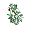
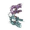
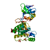
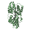
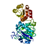
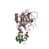
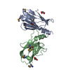
| Title | Structure of reduced C22S OhrR from Xanthamonas Campestris | ||||||
 Components Components | Transcriptional regulator OhrR | ||||||
 Keywords Keywords | TRANSCRIPTION REGULATOR | ||||||
| Function / homology |  Function and homology information Function and homology informationresponse to stress / DNA-binding transcription factor activity / DNA-templated transcription / DNA binding / cytoplasm Similarity search - Function | ||||||
| Biological species |  Xanthomonas campestris (bacteria) Xanthomonas campestris (bacteria) | ||||||
| Method |  X-RAY DIFFRACTION / SAD / MAD / Resolution: 1.9 Å X-RAY DIFFRACTION / SAD / MAD / Resolution: 1.9 Å | ||||||
 Authors Authors | Brennan, R.G. / Newberry, K.J. | ||||||
 Citation Citation | Journal: Mol.Cell / Year: 2007 Title: Structural Mechanism of Organic Hydroperoxide Induction of the Transcription Regulator OhrR. Authors: Newberry, K.J. / Fuangthong, M. / Panmanee, W. / Mongkolsuk, S. / Brennan, R.G. #1: Journal: J.BACTERIOL. / Year: 2006Title: Novel organic hydroperoxide-sensing and responding mechanisms for OhrR, a major bacterial sensor and regulator of organic hydroperoxide stress. Authors: Panmanee, W. / Vattanaviboon, P. / Poole, L.B. / Mongkolsuk, S. | ||||||
| History |
|
- Structure visualization
| Structure viewer | Molecule: MolmilJmol/JSmol |
|---|
- Downloads & links
-Download
| PDBx/mmCIF format | 2pex.cif.gz | 70.5 KB | Display | PDBx/mmCIF format |
|---|---|---|---|---|
| PDB format | pdb2pex.ent.gz | 51.1 KB | Display | PDB format |
| PDBx/mmJSON format | 2pex.json.gz | Tree view | PDBx/mmJSON format | |
| Others |  Other downloads Other downloads |
-Validation report
| Arichive directory | https://data.pdbj.org/pub/pdb/validation_reports/pe/2pexftp://data.pdbj.org/pub/pdb/validation_reports/pe/2pex | HTTPS FTP |
|---|
-Related structure data


-Links
 PDBj
PDBj- Assembly
| Deposited unit | 
| ||||||||
|---|---|---|---|---|---|---|---|---|---|
| 1 |
| ||||||||
| Unit cell |
| ||||||||
| Details | The biological assembly is a dimer |
-Components
| #1: Protein | Mass: 16938.436 Da / Num. of mol.: 2 / Mutation: C22S Source method: isolated from a genetically manipulated source Source: (gene. exp.) Xanthomonas campestris (bacteria) / Gene: ohrR / Plasmid: PET11a / Species (production host): Escherichia coli / Production host: #2: Chemical | ChemComp-FMT /   Mass: 46.025 Da / Num. of mol.: 7 / Source method: obtained synthetically / Formula: CH2O2 Mass: 46.025 Da / Num. of mol.: 7 / Source method: obtained synthetically / Formula: CH2O2#3: Water | ChemComp-HOH / |  Mass: 18.015 Da / Num. of mol.: 175 / Source method: isolated from a natural source / Formula: H2O Mass: 18.015 Da / Num. of mol.: 175 / Source method: isolated from a natural source / Formula: H2O |
|---|
-Experimental details
-Experiment
| Experiment | Method: X-RAY DIFFRACTION / Number of used crystals: 1 |
|---|
- Sample preparation
| Crystal | Density Matthews: 2.42 Å3/Da / Density % sol: 49.27 % |
|---|---|
| Crystal grow | Temperature: 298 K / Method: vapor diffusion, hanging drop Details: 4M sodium formate, VAPOR DIFFUSION, HANGING DROP, temperature 298K |
-Data collection
| Diffraction |
| |||||||||||||||||||||||||||||||||||||||||||||||||||||||||||||||||||||||||||||
|---|---|---|---|---|---|---|---|---|---|---|---|---|---|---|---|---|---|---|---|---|---|---|---|---|---|---|---|---|---|---|---|---|---|---|---|---|---|---|---|---|---|---|---|---|---|---|---|---|---|---|---|---|---|---|---|---|---|---|---|---|---|---|---|---|---|---|---|---|---|---|---|---|---|---|---|---|---|---|
| Diffraction source |
| |||||||||||||||||||||||||||||||||||||||||||||||||||||||||||||||||||||||||||||
| Detector |
| |||||||||||||||||||||||||||||||||||||||||||||||||||||||||||||||||||||||||||||
| Radiation |
| |||||||||||||||||||||||||||||||||||||||||||||||||||||||||||||||||||||||||||||
| Radiation wavelength |
| |||||||||||||||||||||||||||||||||||||||||||||||||||||||||||||||||||||||||||||
| Reflection | Redundancy: 26.69 % / Av σ(I) over netI: 42.1 / Number: 359108 / Rmerge(I) obs: 0.061 / Χ2: 0.94 / D res high: 2.4 Å / D res low: 27.63 Å / Num. obs: 13353 / % possible obs: 99.4 | |||||||||||||||||||||||||||||||||||||||||||||||||||||||||||||||||||||||||||||
| Diffraction reflection shell | ID: 1
| |||||||||||||||||||||||||||||||||||||||||||||||||||||||||||||||||||||||||||||
| Reflection | Resolution: 1.9→25.63 Å / Num. all: 26612 / Num. obs: 26616 / % possible obs: 100 % / Observed criterion σ(F): 0 / Redundancy: 13.79 % / Biso Wilson estimate: 34.1 Å2 / Limit h max: 28 / Limit h min: 0 / Limit k max: 40 / Limit k min: 0 / Limit l max: 40 / Limit l min: 0 / Observed criterion F max: 1160314.12 / Observed criterion F min: 2.5 / Rmerge(I) obs: 0.063 / Χ2: 0.98 / Net I/σ(I): 20.4 / Scaling rejects: 2773 | |||||||||||||||||||||||||||||||||||||||||||||||||||||||||||||||||||||||||||||
| Reflection shell | Diffraction-ID: 1,2 / % possible all: 100
|
-Phasing
| Phasing | Method: MAD | ||||||||||||||||||||||||||||||||||||||||||||||||||||||||
|---|---|---|---|---|---|---|---|---|---|---|---|---|---|---|---|---|---|---|---|---|---|---|---|---|---|---|---|---|---|---|---|---|---|---|---|---|---|---|---|---|---|---|---|---|---|---|---|---|---|---|---|---|---|---|---|---|---|
| Phasing MAD set site |
| ||||||||||||||||||||||||||||||||||||||||||||||||||||||||
| Phasing dm | FOM : 0.71 / FOM acentric: 0.73 / FOM centric: 0.59 / Reflection: 11820 / Reflection acentric: 10091 / Reflection centric: 1729 | ||||||||||||||||||||||||||||||||||||||||||||||||||||||||
| Phasing dm shell |
|
- Processing
| Software |
| ||||||||||||||||||||||||||||||||||||||||||||||||||||||||||||||||||||||||||||||||||||||||||
|---|---|---|---|---|---|---|---|---|---|---|---|---|---|---|---|---|---|---|---|---|---|---|---|---|---|---|---|---|---|---|---|---|---|---|---|---|---|---|---|---|---|---|---|---|---|---|---|---|---|---|---|---|---|---|---|---|---|---|---|---|---|---|---|---|---|---|---|---|---|---|---|---|---|---|---|---|---|---|---|---|---|---|---|---|---|---|---|---|---|---|---|
| Refinement | Method to determine structure: SAD / Resolution: 1.9→25.63 Å / Rfactor Rfree error: 0.005 / Occupancy max: 1 / Occupancy min: 0.5 / Data cutoff high absF: 1160314.125 / Data cutoff low absF: 0 / Isotropic thermal model: RESTRAINED / Cross valid method: THROUGHOUT / σ(F): 0 / Stereochemistry target values: Engh & Huber / Details: BULK SOLVENT MODEL USED
| ||||||||||||||||||||||||||||||||||||||||||||||||||||||||||||||||||||||||||||||||||||||||||
| Solvent computation | Solvent model: FLAT MODEL / Bsol: 49.588 Å2 / ksol: 0.4 e/Å3 | ||||||||||||||||||||||||||||||||||||||||||||||||||||||||||||||||||||||||||||||||||||||||||
| Displacement parameters | Biso max: 68.5 Å2 / Biso mean: 32.8 Å2 / Biso min: 18.04 Å2
| ||||||||||||||||||||||||||||||||||||||||||||||||||||||||||||||||||||||||||||||||||||||||||
| Refine analyze |
| ||||||||||||||||||||||||||||||||||||||||||||||||||||||||||||||||||||||||||||||||||||||||||
| Refinement step | Cycle: LAST / Resolution: 1.9→25.63 Å
| ||||||||||||||||||||||||||||||||||||||||||||||||||||||||||||||||||||||||||||||||||||||||||
| Refine LS restraints |
| ||||||||||||||||||||||||||||||||||||||||||||||||||||||||||||||||||||||||||||||||||||||||||
| LS refinement shell | Refine-ID: X-RAY DIFFRACTION
| ||||||||||||||||||||||||||||||||||||||||||||||||||||||||||||||||||||||||||||||||||||||||||
| Xplor file |
|
