 Movie
Movie Controller

[English] 日本語
 Yorodumi
- PDB-3q63: X-ray crystal structure of protein MLL2253 from Mesorhizobium lot... -
+ Open data

- Basic information
| Entry | Database: PDB / ID: 3q63 | ||||||
|---|---|---|---|---|---|---|---|
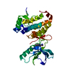
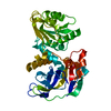
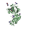
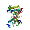
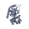
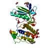
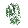
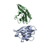
| Title | X-ray crystal structure of protein MLL2253 from Mesorhizobium loti, Northeast Structural Genomics Consortium Target MlR404. | ||||||
 Components Components | Mll2253 protein | ||||||
 Keywords Keywords | Structural Genomics / unknown function / PSI-Biology / Protein Structure Initiative / alpha-beta protein / Unknown / Structure Genomics / Northeast Structural Genomics Consortium / NESG | ||||||
| Function / homology | Activator of Hsp90 ATPase homologue 1-like / Activator of Hsp90 ATPase homolog 1-like protein / START domain / Alpha-D-Glucose-1,6-Bisphosphate; Chain A, domain 4 / START-like domain superfamily / 2-Layer Sandwich / Alpha Beta / Mll2253 protein Function and homology information Function and homology information | ||||||
| Biological species |  Mesorhizobium loti (bacteria) Mesorhizobium loti (bacteria) | ||||||
| Method |  X-RAY DIFFRACTION / SYNCHROTRON / SAD / Resolution: 2 Å X-RAY DIFFRACTION / SYNCHROTRON / SAD / Resolution: 2 Å | ||||||
 Authors Authors | Forouhar, F. / Lew, S. / Seetharaman, J. / Wang, D. / Ciccosanti, C. / Sahdev, S. / Nair, R. / Rost, B. / Acton, T.B. / Xiao, R. ...Forouhar, F. / Lew, S. / Seetharaman, J. / Wang, D. / Ciccosanti, C. / Sahdev, S. / Nair, R. / Rost, B. / Acton, T.B. / Xiao, R. / Everett, J.K. / Montelione, G.T. / Hunt, J.F. / Tong, L. / Northeast Structural Genomics Consortium (NESG) | ||||||
 Citation Citation | Journal: To be Published Title: X-ray crystal structure of protein MLL2253 from Mesorhizobium loti, Northeast Structural Genomics Consortium Target MlR404. Authors: Forouhar, F. / Lew, S. / Seetharaman, J. / Wang, D. / Ciccosanti, C. / Sahdev, S. / Nair, R. / Rost, B. / Acton, T.B. / Xiao, R. / Everett, J.K. / Montelione, G.T. / Hunt, J.F. / Tong, L. | ||||||
| History |
|
- Structure visualization
| Structure viewer | Molecule: MolmilJmol/JSmol |
|---|
- Downloads & links
-Download
| PDBx/mmCIF format | 3q63.cif.gz | 189.9 KB | Display | PDBx/mmCIF format |
|---|---|---|---|---|
| PDB format | pdb3q63.ent.gz | 151.5 KB | Display | PDB format |
| PDBx/mmJSON format | 3q63.json.gz | Tree view | PDBx/mmJSON format | |
| Others |  Other downloads Other downloads |
-Validation report
| Arichive directory | https://data.pdbj.org/pub/pdb/validation_reports/q6/3q63ftp://data.pdbj.org/pub/pdb/validation_reports/q6/3q63 | HTTPS FTP |
|---|
-Related structure data
| Similar structure data | |
|---|---|
| Other databases |


-Links
 PDBj
PDBj- Assembly
| Deposited unit | 
| ||||||||
|---|---|---|---|---|---|---|---|---|---|
| 1 | 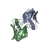
| ||||||||
| 2 | 
| ||||||||
| 3 | 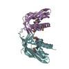
| ||||||||
| Unit cell |
|
-Components
| #1: Protein | Mass: 17757.326 Da / Num. of mol.: 6 Source method: isolated from a genetically manipulated source Source: (gene. exp.) Mesorhizobium loti (bacteria) / Tissue: MAFF303099 / Production host: #2: Water | ChemComp-HOH / |  Mass: 18.015 Da / Num. of mol.: 691 / Source method: isolated from a natural source / Formula: H2O Mass: 18.015 Da / Num. of mol.: 691 / Source method: isolated from a natural source / Formula: H2OHas protein modification | Y | |
|---|
-Experimental details
-Experiment
| Experiment | Method: X-RAY DIFFRACTION / Number of used crystals: 1 |
|---|
- Sample preparation
| Crystal | Density Matthews: 2.4 Å3/Da / Density % sol: 48.7 % |
|---|---|
| Crystal grow | Temperature: 277 K / pH: 5.5 Details: Protein solution: 10 mM Tris (pH 7.5), 100 mM sodium chloride, and 5 mM DTT. Reservoir solution: 100mM BIS-TRIS pH 5.5, 25% PEG 3350, and 200mM Lithium sulfate monohydrate, Microbatch, under ...Details: Protein solution: 10 mM Tris (pH 7.5), 100 mM sodium chloride, and 5 mM DTT. Reservoir solution: 100mM BIS-TRIS pH 5.5, 25% PEG 3350, and 200mM Lithium sulfate monohydrate, Microbatch, under oil, temperature 277K |
-Data collection
| Diffraction | Mean temperature: 100 K |
|---|---|
| Diffraction source | Source: SYNCHROTRON / Site: NSLS  / Beamline: X4A / Wavelength: 0.9789 Å / Beamline: X4A / Wavelength: 0.9789 Å |
| Detector | Type: ADSC QUANTUM 4 / Detector: CCD / Date: Oct 21, 2010 / Details: mirrors |
| Radiation | Monochromator: Si 111 CHANNEL / Protocol: SINGLE WAVELENGTH / Monochromatic (M) / Laue (L): M / Scattering type: x-ray |
| Radiation wavelength | Wavelength: 0.9789 Å / Relative weight: 1 |
| Reflection | Resolution: 2→30 Å / Num. obs: 136539 / % possible obs: 97.8 % / Observed criterion σ(F): 0 / Observed criterion σ(I): 0 / Redundancy: 6 % / Biso Wilson estimate: 12.2 Å2 / Rmerge(I) obs: 0.147 / Rsym value: 0.145 / Net I/σ(I): 14.82 |
| Reflection shell | Resolution: 2→2.07 Å / Redundancy: 4.9 % / Rmerge(I) obs: 0.3 / Mean I/σ(I) obs: 3.9 / Num. unique all: 6853 / Rsym value: 0.315 / % possible all: 96.1 |
- Processing
| Software |
| ||||||||||||||||||||||||||||||||||||||||||||||||||||||||||||
|---|---|---|---|---|---|---|---|---|---|---|---|---|---|---|---|---|---|---|---|---|---|---|---|---|---|---|---|---|---|---|---|---|---|---|---|---|---|---|---|---|---|---|---|---|---|---|---|---|---|---|---|---|---|---|---|---|---|---|---|---|---|
| Refinement | Method to determine structure: SAD / Resolution: 2→19.99 Å / Rfactor Rfree error: 0.002 / Data cutoff high absF: 482644.42 / Data cutoff low absF: 0 / Isotropic thermal model: RESTRAINED / Cross valid method: THROUGHOUT / σ(F): 2 / σ(I): 2 / Stereochemistry target values: Engh & Huber
| ||||||||||||||||||||||||||||||||||||||||||||||||||||||||||||
| Solvent computation | Solvent model: FLAT MODEL / Bsol: 45.6816 Å2 / ksol: 0.4 e/Å3 | ||||||||||||||||||||||||||||||||||||||||||||||||||||||||||||
| Displacement parameters | Biso mean: 25.2 Å2
| ||||||||||||||||||||||||||||||||||||||||||||||||||||||||||||
| Refine analyze |
| ||||||||||||||||||||||||||||||||||||||||||||||||||||||||||||
| Refinement step | Cycle: LAST / Resolution: 2→19.99 Å
| ||||||||||||||||||||||||||||||||||||||||||||||||||||||||||||
| Refine LS restraints |
| ||||||||||||||||||||||||||||||||||||||||||||||||||||||||||||
| LS refinement shell | Resolution: 2→2.07 Å / Rfactor Rfree error: 0.009 / Total num. of bins used: 10
|
