TRANSFERASE / PLK4 / Polo-like kinase 4 / SAK / STK18 / PSI / structural genomics / Protein Structure Initiative / New York SGX Research Center for Structural Genomics / NYSGXRC / ATP-binding / Nucleotide-binding / Phosphoprotein / Polymorphism / Serine/threonine-protein kinase
Function / homology
Function and homology information
de novo centriole assembly involved in multi-ciliated epithelial cell differentiation / procentriole / deuterosome / positive regulation of centriole replication / trophoblast giant cell differentiation / polo kinase / XY body / centriole replication / cleavage furrow / cilium assembly ...de novo centriole assembly involved in multi-ciliated epithelial cell differentiation / procentriole / deuterosome / positive regulation of centriole replication / trophoblast giant cell differentiation / polo kinase / XY body / centriole replication / cleavage furrow / cilium assembly / Loss of Nlp from mitotic centrosomes / Loss of proteins required for interphase microtubule organization from the centrosome / Recruitment of mitotic centrosome proteins and complexes / Recruitment of NuMA to mitotic centrosomes / centriole / Anchoring of the basal body to the plasma membrane / AURKA Activation by TPX2 / Regulation of PLK1 Activity at G2/M Transition / protein phosphorylation / protein serine kinase activity / protein serine/threonine kinase activity / centrosome / nucleolus / ATP binding / identical protein binding / nucleus / cytosol Similarity search - Function
Plk4, C-terminal polo-box domain / Plk4, second cryptic polo-box domain / Plk4, first cryptic polo-box domain / Polo-like Kinase 4 Polo Box 1 / Polo-like Kinase 4 Polo Box 2 / Cryptic Polo-Box 1 (CPB1) domain profile. / Cryptic Polo-Box 2 (CPB2) domain profile. / Serine/threonine-protein kinase, first cryptic polo-box domain superfamily / : / POLO box domain ...Plk4, C-terminal polo-box domain / Plk4, second cryptic polo-box domain / Plk4, first cryptic polo-box domain / Polo-like Kinase 4 Polo Box 1 / Polo-like Kinase 4 Polo Box 2 / Cryptic Polo-Box 1 (CPB1) domain profile. / Cryptic Polo-Box 2 (CPB2) domain profile. / Serine/threonine-protein kinase, first cryptic polo-box domain superfamily / : / POLO box domain / POLO box domain profile. / Tyrosine-protein kinase, active site / Phosphorylase Kinase; domain 1 / Phosphorylase Kinase; domain 1 / Transferase(Phosphotransferase) domain 1 / Transferase(Phosphotransferase); domain 1 / Protein kinase domain / Protein kinase, ATP binding site / Protein kinases ATP-binding region signature. / Protein kinase domain profile. / Protein kinase domain / Protein kinase-like domain superfamily / 2-Layer Sandwich / Orthogonal Bundle / Mainly Alpha / Alpha Beta Similarity search - Domain/homology
In the structure databanks used in Yorodumi, some data are registered as the other names, "COVID-19 virus" and "2019-nCoV". Here are the details of the virus and the list of structure data.
Jan 31, 2019. EMDB accession codes are about to change! (news from PDBe EMDB page)
EMDB accession codes are about to change! (news from PDBe EMDB page)
The allocation of 4 digits for EMDB accession codes will soon come to an end. Whilst these codes will remain in use, new EMDB accession codes will include an additional digit and will expand incrementally as the available range of codes is exhausted. The current 4-digit format prefixed with “EMD-” (i.e. EMD-XXXX) will advance to a 5-digit format (i.e. EMD-XXXXX), and so on. It is currently estimated that the 4-digit codes will be depleted around Spring 2019, at which point the 5-digit format will come into force.
The EM Navigator/Yorodumi systems omit the EMD- prefix.
Related info.:Q: What is EMD? / ID/Accession-code notation in Yorodumi/EM Navigator
Yorodumi is a browser for structure data from EMDB, PDB, SASBDB, etc.
This page is also the successor to EM Navigator detail page, and also detail information page/front-end page for Omokage search.
The word "yorodu" (or yorozu) is an old Japanese word meaning "ten thousand". "mi" (miru) is to see.
Related info.:EMDB / PDB / SASBDB / Comparison of 3 databanks / Yorodumi Search / Aug 31, 2016. New EM Navigator & Yorodumi / Yorodumi Papers / Jmol/JSmol / Function and homology information / Changes in new EM Navigator and Yorodumi
 Movie
Movie Controller


 Open data
 Basic information
 Components
 Keywords
 Function and homology information
 Homo sapiens (human)
 X-RAY DIFFRACTION /
  Authors
 Citation
 Structure visualization
 Downloads & links
 Other downloads


 PDBj
PDBj


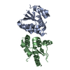
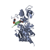
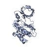
 Assembly


 Mass: 96.063 Da / Num. of mol.: 4 / Source method: obtained synthetically / Formula: SO4
 Mass: 506.196 Da / Num. of mol.: 2 / Source method: obtained synthetically / Formula: C10H17N6O12P3 / Comment: AMP-PNP, energy-carrying molecule analogue*YM
 Mass: 18.015 Da / Num. of mol.: 91 / Source method: isolated from a natural source / Formula: H2O
 Sample preparation
 / Beamline: 31-ID / Wavelength: 0.9794 Å
 Processing