 Movie
Movie Controller

[English] 日本語
 Yorodumi
- PDB-2pa4: Crystal structure of UDP-glucose pyrophosphorylase from Corynebac... -
+ Open data

- Basic information
| Entry | Database: PDB / ID: 2pa4 | ||||||
|---|---|---|---|---|---|---|---|
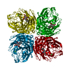
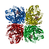
| Title | Crystal structure of UDP-glucose pyrophosphorylase from Corynebacteria glutamicum in complex with magnesium and UDP-glucose | ||||||
 Components Components | UTP-GLUCOSE-1-PHOSPHATE URIDYLYLTRANSFERASE | ||||||
 Keywords Keywords | TRANSFERASE / phosphorylase / nucleotidyltransferase / metabolism | ||||||
| Function / homology | Spore Coat Polysaccharide Biosynthesis Protein SpsA; Chain A / Spore Coat Polysaccharide Biosynthesis Protein SpsA; Chain A / Alpha-Beta Complex / Alpha Beta / URIDINE-5'-DIPHOSPHATE-GLUCOSE / :  Function and homology information Function and homology information | ||||||
| Biological species |  Corynebacterium glutamicum ATCC 13032 (bacteria) Corynebacterium glutamicum ATCC 13032 (bacteria) | ||||||
| Method |  X-RAY DIFFRACTION / SYNCHROTRON / SIR / Resolution: 2 Å X-RAY DIFFRACTION / SYNCHROTRON / SIR / Resolution: 2 Å | ||||||
 Authors Authors | Holden, H.M. / Thoden, J.B. | ||||||
 Citation Citation | Journal: Protein Sci. / Year: 2007 Title: Active site geometry of glucose-1-phosphate uridylyltransferase. Authors: Thoden, J.B. / Holden, H.M. | ||||||
| History |
|
- Structure visualization
| Structure viewer | Molecule: MolmilJmol/JSmol |
|---|
- Downloads & links
-Download
| PDBx/mmCIF format | 2pa4.cif.gz | 252 KB | Display | PDBx/mmCIF format |
|---|---|---|---|---|
| PDB format | pdb2pa4.ent.gz | 202.5 KB | Display | PDB format |
| PDBx/mmJSON format | 2pa4.json.gz | Tree view | PDBx/mmJSON format | |
| Others |  Other downloads Other downloads |
-Validation report
| Arichive directory | https://data.pdbj.org/pub/pdb/validation_reports/pa/2pa4ftp://data.pdbj.org/pub/pdb/validation_reports/pa/2pa4 | HTTPS FTP |
|---|
-Related structure data
| Similar structure data |
|---|


-Links
 PDBj
PDBj
- Assembly
| Deposited unit | 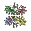
| ||||||||
|---|---|---|---|---|---|---|---|---|---|
| 1 |
| ||||||||
| 2 | 
| ||||||||
| 3 | 
| ||||||||
| Unit cell |
| ||||||||
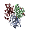
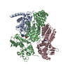
| Details | The biological assembly is a homo-tetramer comprised of chains A, B, C, and D as deposited in the pdb entry |
-Components
| #1: Protein | Mass: 34780.695 Da / Num. of mol.: 4 Source method: isolated from a genetically manipulated source Source: (gene. exp.) Corynebacterium glutamicum ATCC 13032 (bacteria)Species: Corynebacterium glutamicum / Strain: ATCC-13032 / Gene: galU1 / Plasmid: pET31 / Production host: References: UniProt: Q6M6R3, UTP-glucose-1-phosphate uridylyltransferase #2: Chemical | ChemComp-MG /   Mass: 24.305 Da / Num. of mol.: 8 / Source method: obtained synthetically / Formula: Mg Mass: 24.305 Da / Num. of mol.: 8 / Source method: obtained synthetically / Formula: Mg#3: Chemical | ChemComp-UPG /   Mass: 566.302 Da / Num. of mol.: 4 / Source method: obtained synthetically / Formula: C15H24N2O17P2 Mass: 566.302 Da / Num. of mol.: 4 / Source method: obtained synthetically / Formula: C15H24N2O17P2#4: Water | ChemComp-HOH / |  Mass: 18.015 Da / Num. of mol.: 543 / Source method: isolated from a natural source / Formula: H2O Mass: 18.015 Da / Num. of mol.: 543 / Source method: isolated from a natural source / Formula: H2O |
|---|
-Experimental details
-Experiment
| Experiment | Method: X-RAY DIFFRACTION / Number of used crystals: 1 |
|---|
- Sample preparation
| Crystal | Density Matthews: 2.34 Å3/Da / Density % sol: 47.33 % |
|---|---|
| Crystal grow | Temperature: 298 K / Method: vapor diffusion, hanging drop / pH: 6 Details: 23% PEG-3400, 200 mM MgCl2, 200 mM NaCl, 10 mM UDP-glucose, 100 mM MES, pH 6.0, VAPOR DIFFUSION, HANGING DROP, temperature 298K |
-Data collection
| Diffraction | Mean temperature: 100 K |
|---|---|
| Diffraction source | Source: SYNCHROTRON / Site: APS  / Beamline: 19-ID / Wavelength: 0.99848 Å / Beamline: 19-ID / Wavelength: 0.99848 Å |
| Detector | Type: ADSC QUANTUM 315 / Detector: CCD / Date: Aug 6, 2006 / Details: mirrors |
| Radiation | Monochromator: Double-crystal monochromater Si-111 / Protocol: SINGLE WAVELENGTH / Monochromatic (M) / Laue (L): M / Scattering type: x-ray |
| Radiation wavelength | Wavelength: 0.99848 Å / Relative weight: 1 |
| Reflection | Resolution: 2→20 Å / Num. all: 78712 / Num. obs: 78712 / % possible obs: 88.8 % / Observed criterion σ(F): 0 / Observed criterion σ(I): 0 / Redundancy: 6.9 % / Rmerge(I) obs: 0.067 / Rsym value: 0.067 / Net I/σ(I): 33.7 |
| Reflection shell | Resolution: 2→2.07 Å / Redundancy: 3.3 % / Rmerge(I) obs: 0.198 / Mean I/σ(I) obs: 4.5 / Num. unique all: 5571 / Rsym value: 0.198 / % possible all: 83.9 |
- Processing
| Software |
| |||||||||||||||||||||||||
|---|---|---|---|---|---|---|---|---|---|---|---|---|---|---|---|---|---|---|---|---|---|---|---|---|---|---|
| Refinement | Method to determine structure: SIR / Resolution: 2→20 Å / Isotropic thermal model: overall / Cross valid method: THROUGHOUT / σ(F): 0 / σ(I): 0 / Stereochemistry target values: Engh & Huber
| |||||||||||||||||||||||||
| Refinement step | Cycle: LAST / Resolution: 2→20 Å
|
