 Movie
Movie Controller


+ Open data

- Basic information
| Entry | Database: PDB / ID: 2ht7 | ||||||
|---|---|---|---|---|---|---|---|
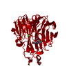
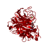
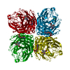
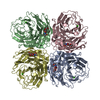
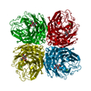
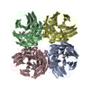
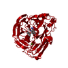
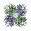
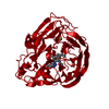
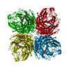
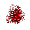
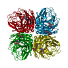
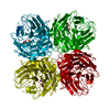
| Title | N8 neuraminidase in open complex with oseltamivir | ||||||
 Components Components | Neuraminidase | ||||||
 Keywords Keywords | HYDROLASE / N8 / neuraminidase / oseltamivir | ||||||
| Function / homology |  Function and homology information Function and homology informationexo-alpha-sialidase / exo-alpha-sialidase activity / viral budding from plasma membrane / carbohydrate metabolic process / host cell plasma membrane / virion membrane / membrane / metal ion binding Similarity search - Function | ||||||
| Biological species |   Influenza A virus Influenza A virus | ||||||
| Method |  X-RAY DIFFRACTION / MOLECULAR REPLACEMENT / Resolution: 2.6 Å X-RAY DIFFRACTION / MOLECULAR REPLACEMENT / Resolution: 2.6 Å | ||||||
 Authors Authors | Russell, R.J. / Haire, L.F. / Stevens, D.J. / Collins, P.J. / Lin, Y.P. / Blackburn, G.M. / Hay, A.J. / Gamblin, S.J. / Skehel, J.J. | ||||||
 Citation Citation | Journal: Nature / Year: 2006 Title: The structure of H5N1 avian influenza neuraminidase suggests new opportunities for drug design. Authors: Russell, R.J. / Haire, L.F. / Stevens, D.J. / Collins, P.J. / Lin, Y.P. / Blackburn, G.M. / Hay, A.J. / Gamblin, S.J. / Skehel, J.J. | ||||||
| History |
|
- Structure visualization
| Structure viewer | Molecule: MolmilJmol/JSmol |
|---|
- Downloads & links
-Download
| PDBx/mmCIF format | 2ht7.cif.gz | 87.3 KB | Display | PDBx/mmCIF format |
|---|---|---|---|---|
| PDB format | pdb2ht7.ent.gz | 65.5 KB | Display | PDB format |
| PDBx/mmJSON format | 2ht7.json.gz | Tree view | PDBx/mmJSON format | |
| Others |  Other downloads Other downloads |
-Validation report
| Arichive directory | https://data.pdbj.org/pub/pdb/validation_reports/ht/2ht7ftp://data.pdbj.org/pub/pdb/validation_reports/ht/2ht7 | HTTPS FTP |
|---|
-Related structure data
| Related structure data |  2ht5C  2ht8C  2htqC 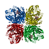 2htrC 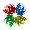 2htuC 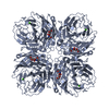 2htvC 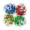 2htwC  2htyC 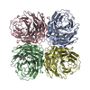 2hu0C 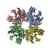 2hu4C C: citing same article ( |
|---|---|
| Similar structure data |


-Links
 PDBj
PDBj

- Assembly
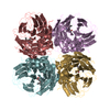
| Deposited unit | 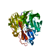
| ||||||||
|---|---|---|---|---|---|---|---|---|---|
| 1 | 
| ||||||||
| Unit cell |
|
-Components
| #1: Protein | Mass: 43157.520 Da / Num. of mol.: 1 / Source method: isolated from a natural source / Source: (natural) Influenza A virus / Genus: Influenzavirus A / References: UniProt: Q07599, exo-alpha-sialidase |
|---|---|
| #2: Chemical | ChemComp-G39 / (  Mass: 284.351 Da / Num. of mol.: 1 / Source method: obtained synthetically / Formula: C14H24N2O4 / Comment: medication*YM Mass: 284.351 Da / Num. of mol.: 1 / Source method: obtained synthetically / Formula: C14H24N2O4 / Comment: medication*YM |
| Has protein modification | Y |
-Experimental details
-Experiment
| Experiment | Method: X-RAY DIFFRACTION / Number of used crystals: 1 |
|---|
- Sample preparation
| Crystal | Density Matthews: 2.63 Å3/Da / Density % sol: 53.26 % |
|---|---|
| Crystal grow | Temperature: 293 K / Method: vapor diffusion, hanging drop / pH: 8 Details: Imidazole, MPD, pH 8, VAPOR DIFFUSION, HANGING DROP, temperature 293K |
-Data collection
| Diffraction | Mean temperature: 100 K |
|---|---|
| Diffraction source | Source: ROTATING ANODE / Type: RIGAKU RU200 / Wavelength: 1.54 Å |
| Detector | Type: RIGAKU RAXIS IIC / Detector: IMAGE PLATE / Date: Feb 2, 2005 |
| Radiation | Protocol: SINGLE WAVELENGTH / Monochromatic (M) / Laue (L): M / Scattering type: x-ray |
| Radiation wavelength | Wavelength: 1.54 Å / Relative weight: 1 |
| Reflection | Resolution: 2.6→30 Å / Num. all: 13800 / Num. obs: 13732 / % possible obs: 99.9 % / Observed criterion σ(F): 0 / Observed criterion σ(I): 0 / Rsym value: 0.147 / Net I/σ(I): 12.6 |
| Reflection shell | Resolution: 2.6→2.7 Å / % possible all: 99.8 |
- Processing
| Software |
| ||||||||||||||||||||
|---|---|---|---|---|---|---|---|---|---|---|---|---|---|---|---|---|---|---|---|---|---|
| Refinement | Method to determine structure: MOLECULAR REPLACEMENT / Resolution: 2.6→30 Å / Cross valid method: THROUGHOUT / σ(F): 0 / Stereochemistry target values: Engh & Huber
| ||||||||||||||||||||
| Refinement step | Cycle: LAST / Resolution: 2.6→30 Å
| ||||||||||||||||||||
| Refine LS restraints |
|
