 Movie
Movie Controller


+ Open data

- Basic information
| Entry | Database: PDB / ID: 2hty | |||||||||
|---|---|---|---|---|---|---|---|---|---|---|
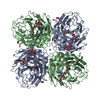
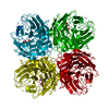
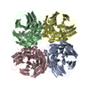
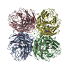
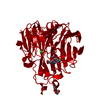
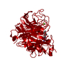
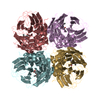
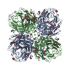
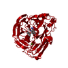
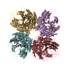
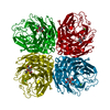
| Title | N1 neuraminidase | |||||||||
 Components Components | Neuraminidase | |||||||||
 Keywords Keywords | HYDROLASE / N1 / neuraminidase | |||||||||
| Function / homology |  Function and homology information Function and homology informationexo-alpha-sialidase / exo-alpha-sialidase activity / viral budding from plasma membrane / carbohydrate metabolic process / host cell plasma membrane / virion membrane / membrane / metal ion binding / identical protein binding Similarity search - Function | |||||||||
| Biological species |   Influenza A virus Influenza A virus | |||||||||
| Method |  X-RAY DIFFRACTION / MOLECULAR REPLACEMENT / Resolution: 2.5 Å X-RAY DIFFRACTION / MOLECULAR REPLACEMENT / Resolution: 2.5 Å | |||||||||
 Authors Authors | Russell, R.J. / Haire, L.F. / Stevens, D.J. / Collins, P.J. / Lin, Y.P. / Blackburn, G.M. / Hay, A.J. / Gamblin, S.J. / Skehel, J.J. | |||||||||
 Citation Citation | Journal: Nature / Year: 2006 Title: The structure of H5N1 avian influenza neuraminidase suggests new opportunities for drug design. Authors: Russell, R.J. / Haire, L.F. / Stevens, D.J. / Collins, P.J. / Lin, Y.P. / Blackburn, G.M. / Hay, A.J. / Gamblin, S.J. / Skehel, J.J. | |||||||||
| History |
|
- Structure visualization
| Structure viewer | Molecule: MolmilJmol/JSmol |
|---|
- Downloads & links
-Download
| PDBx/mmCIF format | 2hty.cif.gz | 595.7 KB | Display | PDBx/mmCIF format |
|---|---|---|---|---|
| PDB format | pdb2hty.ent.gz | 489 KB | Display | PDB format |
| PDBx/mmJSON format | 2hty.json.gz | Tree view | PDBx/mmJSON format | |
| Others |  Other downloads Other downloads |
-Validation report
| Arichive directory | https://data.pdbj.org/pub/pdb/validation_reports/ht/2htyftp://data.pdbj.org/pub/pdb/validation_reports/ht/2hty | HTTPS FTP |
|---|
-Related structure data
| Related structure data |  2ht5C  2ht7C  2ht8C  2htqC 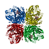 2htrC 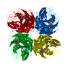 2htuC 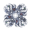 2htvC 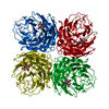 2htwC 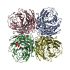 2hu0C 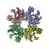 2hu4C C: citing same article ( |
|---|---|
| Similar structure data |


-Links
 PDBj
PDBj


- Assembly
| Deposited unit | 
| ||||||||
|---|---|---|---|---|---|---|---|---|---|
| 1 | 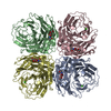
| ||||||||
| 2 | 
| ||||||||
| Unit cell |
|
-Components
| #1: Protein | Mass: 42429.180 Da / Num. of mol.: 8 / Mutation: H253Y / Source method: isolated from a natural source / Source: (natural) Influenza A virus / Genus: Influenzavirus A / References: UniProt: Q6DPL2#2: Sugar | ChemComp-NDG /   Type: D-saccharide, alpha linking / Mass: 221.208 Da / Num. of mol.: 7 Type: D-saccharide, alpha linking / Mass: 221.208 Da / Num. of mol.: 7Source method: isolated from a genetically manipulated source Formula: C8H15NO6 #3: Chemical | ChemComp-CA /   Mass: 40.078 Da / Num. of mol.: 8 / Source method: obtained synthetically / Formula: Ca Mass: 40.078 Da / Num. of mol.: 8 / Source method: obtained synthetically / Formula: Ca#4: Sugar | ChemComp-NAG / |   Type: D-saccharide, beta linking / Mass: 221.208 Da / Num. of mol.: 1 Type: D-saccharide, beta linking / Mass: 221.208 Da / Num. of mol.: 1Source method: isolated from a genetically manipulated source Formula: C8H15NO6 #5: Water | ChemComp-HOH / |  Mass: 18.015 Da / Num. of mol.: 634 / Source method: isolated from a natural source / Formula: H2O Mass: 18.015 Da / Num. of mol.: 634 / Source method: isolated from a natural source / Formula: H2OHas protein modification | Y | |
|---|
-Experimental details
-Experiment
| Experiment | Method: X-RAY DIFFRACTION / Number of used crystals: 1 |
|---|
- Sample preparation
| Crystal | Density Matthews: 3.13 Å3/Da / Density % sol: 60.73 % |
|---|
-Data collection
| Diffraction | Mean temperature: 100 K |
|---|---|
| Diffraction source | Source: ROTATING ANODE / Type: RIGAKU / Wavelength: 1.54 Å |
| Detector | Type: RIGAKU RAXIS IV / Detector: IMAGE PLATE / Date: Mar 3, 2006 |
| Radiation | Protocol: SINGLE WAVELENGTH / Monochromatic (M) / Laue (L): M / Scattering type: x-ray |
| Radiation wavelength | Wavelength: 1.54 Å / Relative weight: 1 |
| Reflection | Resolution: 2.5→30 Å / Num. all: 146515 / Num. obs: 143352 / % possible obs: 98 % / Observed criterion σ(F): 0 / Observed criterion σ(I): 0 / Rsym value: 0.119 / Net I/σ(I): 7.1 |
- Processing
| Software |
| ||||||||||||||||||||
|---|---|---|---|---|---|---|---|---|---|---|---|---|---|---|---|---|---|---|---|---|---|
| Refinement | Method to determine structure: MOLECULAR REPLACEMENT / Resolution: 2.5→30 Å / Cross valid method: THROUGHOUT / σ(F): 0 / Stereochemistry target values: Engh & Huber
| ||||||||||||||||||||
| Refinement step | Cycle: LAST / Resolution: 2.5→30 Å
|
