 Movie
Movie Controller

[English] 日本語
 Yorodumi
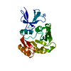
- PDB-2p7g: X-ray Structure of Estrogen Related Receptor g in complex with Bi... -
+ Open data

- Basic information
| Entry | Database: PDB / ID: 2p7g | ||||||
|---|---|---|---|---|---|---|---|
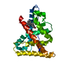
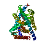
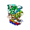
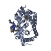
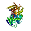
| Title | X-ray Structure of Estrogen Related Receptor g in complex with Bisphenol A. | ||||||
 Components Components | Estrogen-related receptor gamma | ||||||
 Keywords Keywords | HORMONE RECEPTOR / three layered alpha helical sandwich | ||||||
| Function / homology |  Function and homology information Function and homology informationAF-2 domain binding / nuclear steroid receptor activity / estrogen response element binding / retinoic acid receptor signaling pathway / steroid binding / Nuclear Receptor transcription pathway / nuclear receptor activity / sequence-specific double-stranded DNA binding / positive regulation of cold-induced thermogenesis / DNA-binding transcription activator activity, RNA polymerase II-specific ...AF-2 domain binding / nuclear steroid receptor activity / estrogen response element binding / retinoic acid receptor signaling pathway / steroid binding / Nuclear Receptor transcription pathway / nuclear receptor activity / sequence-specific double-stranded DNA binding / positive regulation of cold-induced thermogenesis / DNA-binding transcription activator activity, RNA polymerase II-specific / DNA-binding transcription factor activity, RNA polymerase II-specific / regulation of transcription by RNA polymerase II / regulation of DNA-templated transcription / chromatin / positive regulation of transcription by RNA polymerase II / zinc ion binding / nucleoplasm / identical protein binding / nucleus Similarity search - Function | ||||||
| Biological species |  Homo sapiens (human) Homo sapiens (human) | ||||||
| Method |  X-RAY DIFFRACTION / MOLECULAR REPLACEMENT / Resolution: 2.1 Å X-RAY DIFFRACTION / MOLECULAR REPLACEMENT / Resolution: 2.1 Å | ||||||
 Authors Authors | Abad, M.C. | ||||||
 Citation Citation | Journal: J.Steroid Biochem.Mol.Biol. / Year: 2008 Title: Structural determination of estrogen-related receptor gamma in the presence of phenol derivative compounds. Authors: Abad, M.C. / Askari, H. / O'Neill, J. / Klinger, A.L. / Milligan, C. / Lewandowski, F. / Springer, B. / Spurlino, J. / Rentzeperis, D. | ||||||
| History |
|
- Structure visualization
| Structure viewer | Molecule: MolmilJmol/JSmol |
|---|
- Downloads & links
-Download
| PDBx/mmCIF format | 2p7g.cif.gz | 62.6 KB | Display | PDBx/mmCIF format |
|---|---|---|---|---|
| PDB format | pdb2p7g.ent.gz | 44.1 KB | Display | PDB format |
| PDBx/mmJSON format | 2p7g.json.gz | Tree view | PDBx/mmJSON format | |
| Others |  Other downloads Other downloads |
-Validation report
| Arichive directory | https://data.pdbj.org/pub/pdb/validation_reports/p7/2p7gftp://data.pdbj.org/pub/pdb/validation_reports/p7/2p7g | HTTPS FTP |
|---|
-Related structure data
| Related structure data |  2p7aC  2p7zC  1kv6S C: citing same article ( S: Starting model for refinement |
|---|---|
| Similar structure data |


-Links
 PDBj
PDBj

- Assembly
| Deposited unit | 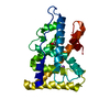
| ||||||||
|---|---|---|---|---|---|---|---|---|---|
| 1 |
| ||||||||
| Unit cell |
|
-Components
| #1: Protein | Mass: 28386.941 Da / Num. of mol.: 1 Source method: isolated from a genetically manipulated source Source: (gene. exp.) Homo sapiens (human) / Gene: ESRRG, ERR3, ERRG2, KIAA0832, NR3B3 / Plasmid: pET28a / Production host:  |
|---|---|
| #2: Chemical | ChemComp-NA /   Mass: 22.990 Da / Num. of mol.: 1 / Source method: obtained synthetically / Formula: Na Mass: 22.990 Da / Num. of mol.: 1 / Source method: obtained synthetically / Formula: Na |
| #3: Chemical | ChemComp-2OH / 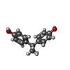  Mass: 228.286 Da / Num. of mol.: 1 / Source method: obtained synthetically / Formula: C15H16O2 Mass: 228.286 Da / Num. of mol.: 1 / Source method: obtained synthetically / Formula: C15H16O2 |
| #4: Water | ChemComp-HOH /  Mass: 18.015 Da / Num. of mol.: 172 / Source method: isolated from a natural source / Formula: H2O Mass: 18.015 Da / Num. of mol.: 172 / Source method: isolated from a natural source / Formula: H2O |
-Experimental details
-Experiment
| Experiment | Method: X-RAY DIFFRACTION / Number of used crystals: 1 |
|---|
- Sample preparation
| Crystal | Density Matthews: 2.47 Å3/Da / Density % sol: 50.11 % |
|---|---|
| Crystal grow | Temperature: 277 K / Method: vapor diffusion, hanging drop / pH: 8.5 Details: 22.6% polyethylene glycol 4000, 0.1M Tris, pH 8.5 and 0.2M Sodium Acetate, VAPOR DIFFUSION, HANGING DROP, temperature 277K |
-Data collection
| Diffraction | Mean temperature: 200 K |
|---|---|
| Diffraction source | Source: ROTATING ANODE / Wavelength: 1.5418 |
| Detector | Type: BRUKER SMART 6000 / Detector: CCD / Date: Aug 6, 2003 / Details: mirrors |
| Radiation | Protocol: SINGLE WAVELENGTH / Monochromatic (M) / Laue (L): M / Scattering type: x-ray |
| Radiation wavelength | Wavelength: 1.5418 Å / Relative weight: 1 |
| Reflection | Resolution: 2.1→68.5 Å / Num. obs: 17455 / Redundancy: 2.5 % / Biso Wilson estimate: 18 Å2 / Rmerge(I) obs: 0.048 / Net I/σ(I): 11.7 |
| Reflection shell | Resolution: 2.1→2.15 Å / Redundancy: 2.5 % / Rmerge(I) obs: 0.14 / Mean I/σ(I) obs: 4.3 / Num. unique all: 3595 / Rsym value: 0.133 |
- Processing
| Software |
| |||||||||||||||||||||||||
|---|---|---|---|---|---|---|---|---|---|---|---|---|---|---|---|---|---|---|---|---|---|---|---|---|---|---|
| Refinement | Method to determine structure: MOLECULAR REPLACEMENT Starting model: pdb entry 1KV6 Resolution: 2.1→68.5 Å / Isotropic thermal model: anisotropic / σ(F): 0 / σ(I): 0 / Stereochemistry target values: Engh & Huber
| |||||||||||||||||||||||||
| Refinement step | Cycle: LAST / Resolution: 2.1→68.5 Å
| |||||||||||||||||||||||||
| Refine LS restraints |
|
