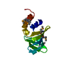
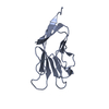
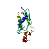
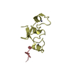
- PDB-2p3h: Crystal structure of the CorC_HlyC domain of a putative Corynebac... -
+
Open data
ID or keywords:
Loading...
-
Basic information
Entry
Database: PDB / ID: 2p3h
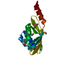
Title
Crystal structure of the CorC_HlyC domain of a putative Corynebacterium glutamicum hemolysin
Components
Uncharacterized CBS domain-containing protein
Keywords
TRANSPORT PROTEIN / structural genomics / CorC_HlyC / pfam03471 / Putative transport protein / Transporter associated domain / Corynebacterium glutamicum / PSI-2 / Protein Structure Initiative / Midwest Center for Structural Genomics / MCSG
BIOMOLECULE: 1 THIS ENTRY CONTAINS THE CRYSTALLOGRAPHIC ASYMMETRIC UNIT WHICH CONSISTS OF 1 ... BIOMOLECULE: 1 THIS ENTRY CONTAINS THE CRYSTALLOGRAPHIC ASYMMETRIC UNIT WHICH CONSISTS OF 1 CHAIN(S). AUTHORS STATE THAT THE BIOLOGICAL UNIT OF THIS POLYPEPTIDE IS UNKNOWN. THE CRYSTALLIZED DOMAIN IS PART OF MUCH LARGER PROTEIN.
Type: CUSTOM-MADE / Detector: CCD / Date: Feb 23, 2007
Radiation
Monochromator: SAGITALLY FOCUSED Si(111) / Protocol: SINGLE WAVELENGTH / Monochromatic (M) / Laue (L): M / Scattering type: x-ray
Radiation wavelength
Wavelength: 0.91946 Å / Relative weight: 1
Reflection
Redundancy: 7.7 % / Av σ(I) over netI: 13.3 / Number: 104059 / Rmerge(I) obs: 0.064 / Χ2: 2.18 / D res high: 1.75 Å / D res low: 50 Å / Num. obs: 13454 / % possible obs: 97.4
In the structure databanks used in Yorodumi, some data are registered as the other names, "COVID-19 virus" and "2019-nCoV". Here are the details of the virus and the list of structure data.
Jan 31, 2019. EMDB accession codes are about to change! (news from PDBe EMDB page)
EMDB accession codes are about to change! (news from PDBe EMDB page)
The allocation of 4 digits for EMDB accession codes will soon come to an end. Whilst these codes will remain in use, new EMDB accession codes will include an additional digit and will expand incrementally as the available range of codes is exhausted. The current 4-digit format prefixed with “EMD-” (i.e. EMD-XXXX) will advance to a 5-digit format (i.e. EMD-XXXXX), and so on. It is currently estimated that the 4-digit codes will be depleted around Spring 2019, at which point the 5-digit format will come into force.
The EM Navigator/Yorodumi systems omit the EMD- prefix.
Related info.:Q: What is EMD? / ID/Accession-code notation in Yorodumi/EM Navigator
Yorodumi is a browser for structure data from EMDB, PDB, SASBDB, etc.
This page is also the successor to EM Navigator detail page, and also detail information page/front-end page for Omokage search.
The word "yorodu" (or yorozu) is an old Japanese word meaning "ten thousand". "mi" (miru) is to see.
Related info.:EMDB / PDB / SASBDB / Comparison of 3 databanks / Yorodumi Search / Aug 31, 2016. New EM Navigator & Yorodumi / Yorodumi Papers / Jmol/JSmol / Function and homology information / Changes in new EM Navigator and Yorodumi
 Movie
Movie Controller

 Yorodumi
 Open data
 Basic information
 Components
 Keywords
 Function and homology information
 Corynebacterium glutamicum (bacteria)
 X-RAY DIFFRACTION /
  Authors
 Citation
 Structure visualization
 Downloads & links
 Other downloads
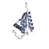

 PDBj
PDBj


 Assembly

 Mass: 79.904 Da / Num. of mol.: 9 / Source method: obtained synthetically / Formula: Br
 Mass: 62.068 Da / Num. of mol.: 3 / Source method: obtained synthetically / Formula: C2H6O2
 Mass: 18.015 Da / Num. of mol.: 172 / Source method: isolated from a natural source / Formula: H2O
 Sample preparation
 / Beamline: 19-BM / Wavelength: 0.91946 Å
 Processing