 Movie
Movie Controller

[English] 日本語
 Yorodumi
- PDB-2p1v: Crystal structure of the ligand binding domain of the retinoid X ... -
+ Open data

- Basic information
| Entry | Database: PDB / ID: 2p1v | ||||||
|---|---|---|---|---|---|---|---|
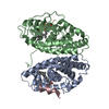
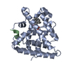
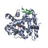
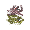
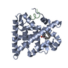
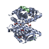
| Title | Crystal structure of the ligand binding domain of the retinoid X receptor alpha in complex with 3-(2'-propoxy)-tetrahydronaphtyl cinnamic acid and a fragment of the coactivator TIF-2 | ||||||
 Components Components |
| ||||||
 Keywords Keywords | HORMONE RECEPTOR / protein-ligand complex | ||||||
| Function / homology |  Function and homology information Function and homology informationretinoic acid-responsive element binding / NR1H2 & NR1H3 regulate gene expression linked to triglyceride lipolysis in adipose / NR1H2 & NR1H3 regulate gene expression linked to gluconeogenesis / NR1H2 & NR1H3 regulate gene expression to limit cholesterol uptake / positive regulation of thyroid hormone receptor signaling pathway / NR1H2 & NR1H3 regulate gene expression linked to lipogenesis / Carnitine shuttle / retinoic acid binding / TGFBR3 expression / positive regulation of vitamin D receptor signaling pathway ...retinoic acid-responsive element binding / NR1H2 & NR1H3 regulate gene expression linked to triglyceride lipolysis in adipose / NR1H2 & NR1H3 regulate gene expression linked to gluconeogenesis / NR1H2 & NR1H3 regulate gene expression to limit cholesterol uptake / positive regulation of thyroid hormone receptor signaling pathway / NR1H2 & NR1H3 regulate gene expression linked to lipogenesis / Carnitine shuttle / retinoic acid binding / TGFBR3 expression / positive regulation of vitamin D receptor signaling pathway / nuclear vitamin D receptor binding / Signaling by Retinoic Acid / RNA polymerase II intronic transcription regulatory region sequence-specific DNA binding / DNA binding domain binding / NR1H2 & NR1H3 regulate gene expression to control bile acid homeostasis / LBD domain binding / locomotor rhythm / aryl hydrocarbon receptor binding / nuclear steroid receptor activity / cellular response to Thyroglobulin triiodothyronine / regulation of lipid metabolic process / regulation of glucose metabolic process / Synthesis of bile acids and bile salts / positive regulation of cholesterol efflux / Synthesis of bile acids and bile salts via 27-hydroxycholesterol / Endogenous sterols / Synthesis of bile acids and bile salts via 7alpha-hydroxycholesterol / response to retinoic acid / positive regulation of bone mineralization / cellular response to hormone stimulus / Recycling of bile acids and salts / Transcriptional regulation of brown and beige adipocyte differentiation by EBF2 / transcription regulator inhibitor activity / retinoic acid receptor signaling pathway / NR1H3 & NR1H2 regulate gene expression linked to cholesterol transport and efflux / hormone-mediated signaling pathway / : / positive regulation of adipose tissue development / Regulation of lipid metabolism by PPARalpha / peroxisome proliferator activated receptor signaling pathway / regulation of cellular response to insulin stimulus / peptide binding / BMAL1:CLOCK,NPAS2 activates circadian expression / SUMOylation of transcription cofactors / Activation of gene expression by SREBF (SREBP) / response to progesterone / nuclear receptor binding / transcription coregulator binding / negative regulation of smoothened signaling pathway / RNA polymerase II transcription regulatory region sequence-specific DNA binding / SUMOylation of intracellular receptors / circadian regulation of gene expression / mRNA transcription by RNA polymerase II / Heme signaling / Transcriptional activation of mitochondrial biogenesis / PPARA activates gene expression / Cytoprotection by HMOX1 / Activated PKN1 stimulates transcription of AR (androgen receptor) regulated genes KLK2 and KLK3 / Nuclear Receptor transcription pathway / Transcriptional regulation of white adipocyte differentiation / RNA polymerase II transcription regulator complex / Activation of anterior HOX genes in hindbrain development during early embryogenesis / nuclear receptor activity / Transcriptional regulation of granulopoiesis / sequence-specific double-stranded DNA binding / : / nervous system development / HATs acetylate histones / MLL4 and MLL3 complexes regulate expression of PPARG target genes in adipogenesis and hepatic steatosis / double-stranded DNA binding / transcription regulator complex / Estrogen-dependent gene expression / sequence-specific DNA binding / DNA-binding transcription factor activity, RNA polymerase II-specific / cell differentiation / transcription coactivator activity / receptor complex / transcription cis-regulatory region binding / protein dimerization activity / nuclear body / RNA polymerase II cis-regulatory region sequence-specific DNA binding / DNA-binding transcription factor activity / protein domain specific binding / chromatin binding / regulation of DNA-templated transcription / chromatin / positive regulation of DNA-templated transcription / enzyme binding / negative regulation of transcription by RNA polymerase II / positive regulation of transcription by RNA polymerase II / protein-containing complex / mitochondrion / zinc ion binding / nucleoplasm / identical protein binding / nucleus / cytoplasm / cytosol Similarity search - Function | ||||||
| Biological species |  Homo sapiens (human) Homo sapiens (human) | ||||||
| Method |  X-RAY DIFFRACTION / SYNCHROTRON / MOLECULAR REPLACEMENT / Resolution: 2.2 Å X-RAY DIFFRACTION / SYNCHROTRON / MOLECULAR REPLACEMENT / Resolution: 2.2 Å | ||||||
 Authors Authors | Bourguet, W. / Nahoum, V. | ||||||
 Citation Citation | Journal: Proc.Natl.Acad.Sci.Usa / Year: 2007 Title: Modulators of the structural dynamics of the retinoid X receptor to reveal receptor function. Authors: Nahoum, V. / Perez, E. / Germain, P. / Rodriguez-Barrios, F. / Manzo, F. / Kammerer, S. / Lemaire, G. / Hirsch, O. / Royer, C.A. / Gronemeyer, H. / de Lera, A.R. / Bourguet, W. | ||||||
| History |
|
- Structure visualization
| Structure viewer | Molecule: MolmilJmol/JSmol |
|---|
- Downloads & links
-Download
| PDBx/mmCIF format | 2p1v.cif.gz | 60.6 KB | Display | PDBx/mmCIF format |
|---|---|---|---|---|
| PDB format | pdb2p1v.ent.gz | 42.6 KB | Display | PDB format |
| PDBx/mmJSON format | 2p1v.json.gz | Tree view | PDBx/mmJSON format | |
| Others |  Other downloads Other downloads |
-Validation report
| Summary document | 2p1v_validation.pdf.gz | 739.3 KB | Display | wwPDB validaton report |
|---|---|---|---|---|
| Full document | 2p1v_full_validation.pdf.gz | 739.7 KB | Display | |
| Data in XML | 2p1v_validation.xml.gz | 11.1 KB | Display | |
| Data in CIF | 2p1v_validation.cif.gz | 14.8 KB | Display | |
| Arichive directory | https://data.pdbj.org/pub/pdb/validation_reports/p1/2p1vftp://data.pdbj.org/pub/pdb/validation_reports/p1/2p1v | HTTPS FTP |
-Related structure data
| Related structure data |  2p1tC 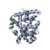 2p1uC  1mvcS S: Starting model for refinement C: citing same article ( |
|---|---|
| Similar structure data |


-Links
 PDBj
PDBj


- Assembly
| Deposited unit | 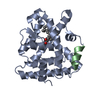
| ||||||||
|---|---|---|---|---|---|---|---|---|---|
| 1 | 
| ||||||||
| Unit cell |
| ||||||||
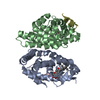
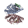
| Details | The biological assembly is a homodimer generated from the monomer in the asymmetric unit by the transformation matrix: 0.0 -1.0 0.0 -1.0 0.0 0.0 0.0 0.0 -1.0 0.0 0.0 0.5 |
-Components
| #1: Protein | Mass: 26856.039 Da / Num. of mol.: 1 / Fragment: ligand binding domain (residues 223-462) Source method: isolated from a genetically manipulated source Source: (gene. exp.) Homo sapiens (human) / Gene: RXRA, NR2B1 / Plasmid: PET15B / Species (production host): Escherichia coli / Production host:  |
|---|---|
| #2: Protein/peptide | Mass: 1579.866 Da / Num. of mol.: 1 Fragment: nuclear receptor interaction motif 2 (residues 686-698) Source method: obtained synthetically Details: This sequence occurs naturally in humans. The peptide has been synthesized by automatic chemical synthesis. References: UniProt: Q15596 |
| #3: Chemical | ChemComp-5TN / (  Mass: 408.530 Da / Num. of mol.: 1 / Source method: obtained synthetically / Formula: C26H32O4 Mass: 408.530 Da / Num. of mol.: 1 / Source method: obtained synthetically / Formula: C26H32O4 |
| #4: Water | ChemComp-HOH /  Mass: 18.015 Da / Num. of mol.: 74 / Source method: isolated from a natural source / Formula: H2O Mass: 18.015 Da / Num. of mol.: 74 / Source method: isolated from a natural source / Formula: H2O |
-Experimental details
-Experiment
| Experiment | Method: X-RAY DIFFRACTION / Number of used crystals: 1 |
|---|
- Sample preparation
| Crystal | Density Matthews: 2.05 Å3/Da / Density % sol: 39.88 % |
|---|---|
| Crystal grow | Temperature: 298 K / Method: vapor diffusion, hanging drop / pH: 7.5 Details: 24% PEG 4000, 0.1M Tris, pH 7.5, VAPOR DIFFUSION, HANGING DROP, temperature 298K |
-Data collection
| Diffraction | Mean temperature: 100 K |
|---|---|
| Diffraction source | Source: SYNCHROTRON / Site: ESRF  / Beamline: ID14-4 / Wavelength: 0.975 / Beamline: ID14-4 / Wavelength: 0.975 |
| Detector | Type: ADSC QUANTUM 315 / Detector: CCD / Date: Nov 12, 2006 / Details: Toroidal mirror |
| Radiation | Monochromator: Double crystal, Si(111) or Si(311) / Protocol: SINGLE WAVELENGTH / Monochromatic (M) / Laue (L): M / Scattering type: x-ray |
| Radiation wavelength | Wavelength: 0.975 Å / Relative weight: 1 |
| Reflection | Resolution: 2.2→35.4 Å / Num. all: 12643 / Num. obs: 11503 / % possible obs: 91.6 % / Observed criterion σ(F): 0 / Observed criterion σ(I): 0 / Redundancy: 10.2 % / Biso Wilson estimate: 32.18 Å2 / Rmerge(I) obs: 0.094 / Rsym value: 0.094 / Net I/σ(I): 5.7 |
| Reflection shell | Resolution: 2.2→2.32 Å / Redundancy: 10.6 % / Rmerge(I) obs: 0.235 / Mean I/σ(I) obs: 2.9 / Num. measured all: 16418 / Num. unique all: 1549 / Rsym value: 0.235 / % possible all: 87.1 |
- Processing
| Software |
| ||||||||||||||||||||||||||||||||||||||||||||||||||||||||||||||||||||||||||||||||||||||||||
|---|---|---|---|---|---|---|---|---|---|---|---|---|---|---|---|---|---|---|---|---|---|---|---|---|---|---|---|---|---|---|---|---|---|---|---|---|---|---|---|---|---|---|---|---|---|---|---|---|---|---|---|---|---|---|---|---|---|---|---|---|---|---|---|---|---|---|---|---|---|---|---|---|---|---|---|---|---|---|---|---|---|---|---|---|---|---|---|---|---|---|---|
| Refinement | Method to determine structure: MOLECULAR REPLACEMENT Starting model: PDB entry 1mvc Resolution: 2.2→35.4 Å / Cor.coef. Fo:Fc: 0.938 / Cor.coef. Fo:Fc free: 0.922 / SU B: 11.235 / SU ML: 0.135 / Isotropic thermal model: Isotropic / Cross valid method: THROUGHOUT / σ(F): 0 / ESU R: 0.329 / ESU R Free: 0.221 / Stereochemistry target values: MAXIMUM LIKELIHOOD / Details: HYDROGENS HAVE BEEN ADDED IN THE RIDING POSITIONS
| ||||||||||||||||||||||||||||||||||||||||||||||||||||||||||||||||||||||||||||||||||||||||||
| Solvent computation | Ion probe radii: 0.8 Å / Shrinkage radii: 0.8 Å / VDW probe radii: 1.4 Å / Solvent model: MASK | ||||||||||||||||||||||||||||||||||||||||||||||||||||||||||||||||||||||||||||||||||||||||||
| Displacement parameters | Biso mean: 29.102 Å2
| ||||||||||||||||||||||||||||||||||||||||||||||||||||||||||||||||||||||||||||||||||||||||||
| Refinement step | Cycle: LAST / Resolution: 2.2→35.4 Å
| ||||||||||||||||||||||||||||||||||||||||||||||||||||||||||||||||||||||||||||||||||||||||||
| Refine LS restraints |
| ||||||||||||||||||||||||||||||||||||||||||||||||||||||||||||||||||||||||||||||||||||||||||
| LS refinement shell | Resolution: 2.2→2.257 Å / Total num. of bins used: 20
|
