 Movie
Movie Controller

[English] 日本語
 Yorodumi
- PDB-2ouh: Crystal structure of the Thrombospondin-1 N-terminal domain in co... -
+ Open data

- Basic information
| Entry | Database: PDB / ID: 2ouh | ||||||
|---|---|---|---|---|---|---|---|
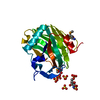
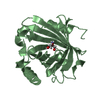
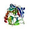
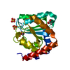
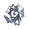
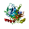
| Title | Crystal structure of the Thrombospondin-1 N-terminal domain in complex with fractionated Heparin DP10 | ||||||
 Components Components | Thrombospondin-1 | ||||||
 Keywords Keywords | CELL ADHESION / TSP-1 / TSPN-1 / HBD / Fractionated Heparin / DP10 | ||||||
| Function / homology |  Function and homology information Function and homology informationnegative regulation of antigen processing and presentation of peptide or polysaccharide antigen via MHC class II / negative regulation of nitric oxide-cGMP mediated signal transduction / negative regulation of dendritic cell antigen processing and presentation / : / collagen V binding / negative regulation of sprouting angiogenesis / chronic inflammatory response / negative regulation of endothelial cell chemotaxis / Defective B3GALTL causes PpS / O-glycosylation of TSR domain-containing proteins ...negative regulation of antigen processing and presentation of peptide or polysaccharide antigen via MHC class II / negative regulation of nitric oxide-cGMP mediated signal transduction / negative regulation of dendritic cell antigen processing and presentation / : / collagen V binding / negative regulation of sprouting angiogenesis / chronic inflammatory response / negative regulation of endothelial cell chemotaxis / Defective B3GALTL causes PpS / O-glycosylation of TSR domain-containing proteins / positive regulation of extrinsic apoptotic signaling pathway via death domain receptors / negative regulation of fibroblast growth factor receptor signaling pathway / negative regulation of long-chain fatty acid import across plasma membrane / negative regulation of blood vessel endothelial cell proliferation involved in sprouting angiogenesis / positive regulation of transforming growth factor beta1 production / fibrinogen complex / engulfment of apoptotic cell / low-density lipoprotein particle binding / negative regulation of interleukin-12 production / negative regulation of focal adhesion assembly / Signaling by PDGF / platelet alpha granule / negative regulation of plasminogen activation / fibrinogen binding / negative regulation of cell migration involved in sprouting angiogenesis / positive regulation of chemotaxis / positive regulation of macrophage activation / transforming growth factor beta binding / sprouting angiogenesis / proteoglycan binding / negative regulation of endothelial cell migration / positive regulation of fibroblast migration / negative regulation of receptor guanylyl cyclase signaling pathway / extracellular matrix structural constituent / Syndecan interactions / phosphatidylserine binding / negative regulation of interleukin-10 production / endopeptidase inhibitor activity / response to testosterone / negative regulation of endothelial cell proliferation / positive regulation of transforming growth factor beta receptor signaling pathway / positive regulation of macrophage chemotaxis / fibronectin binding / negative regulation of blood vessel endothelial cell migration / fibroblast growth factor binding / negative regulation of fibrinolysis / behavioral response to pain / negative regulation of cell-matrix adhesion / blood coagulation, fibrin clot formation / negative regulation of tumor necrosis factor production / positive regulation of phosphorylation / positive regulation of endothelial cell apoptotic process / response to magnesium ion / positive regulation of blood vessel endothelial cell migration / response to unfolded protein / Integrin cell surface interactions / response to mechanical stimulus / response to glucose / nitric oxide-cGMP-mediated signaling / response to progesterone / laminin binding / positive regulation of endothelial cell migration / secretory granule / positive regulation of smooth muscle cell proliferation / platelet alpha granule lumen / response to endoplasmic reticulum stress / negative regulation of angiogenesis / negative regulation of extrinsic apoptotic signaling pathway / sarcoplasmic reticulum / positive regulation of translation / RUNX1 regulates genes involved in megakaryocyte differentiation and platelet function / response to calcium ion / integrin binding / cellular response to growth factor stimulus / cellular response to tumor necrosis factor / positive regulation of reactive oxygen species metabolic process / positive regulation of angiogenesis / positive regulation of tumor necrosis factor production / Platelet degranulation / cell migration / heparin binding / cellular response to heat / extracellular matrix / protease binding / response to hypoxia / positive regulation of MAPK cascade / positive regulation of phosphatidylinositol 3-kinase/protein kinase B signal transduction / cell adhesion / immune response / positive regulation of cell migration / response to xenobiotic stimulus / endoplasmic reticulum lumen / inflammatory response / external side of plasma membrane / negative regulation of cell population proliferation / apoptotic process / calcium ion binding / positive regulation of cell population proliferation / negative regulation of apoptotic process / cell surface Similarity search - Function | ||||||
| Biological species |  Homo sapiens (human) Homo sapiens (human) | ||||||
| Method |  X-RAY DIFFRACTION / SYNCHROTRON / MOLECULAR REPLACEMENT / Resolution: 2.4 Å X-RAY DIFFRACTION / SYNCHROTRON / MOLECULAR REPLACEMENT / Resolution: 2.4 Å | ||||||
 Authors Authors | Tan, K. / Joachimiak, A. / Wang, J. / Lawler, J. | ||||||
 Citation Citation | Journal: J.Biol.Chem. / Year: 2008 Title: Heparin-induced cis- and trans-Dimerization Modes of the Thrombospondin-1 N-terminal Domain. Authors: Tan, K. / Duquette, M. / Liu, J.H. / Shanmugasundaram, K. / Joachimiak, A. / Gallagher, J.T. / Rigby, A.C. / Wang, J.H. / Lawler, J. | ||||||
| History |
|
- Structure visualization
| Structure viewer | Molecule: MolmilJmol/JSmol |
|---|
- Downloads & links
-Download
| PDBx/mmCIF format | 2ouh.cif.gz | 94.3 KB | Display | PDBx/mmCIF format |
|---|---|---|---|---|
| PDB format | pdb2ouh.ent.gz | 71.6 KB | Display | PDB format |
| PDBx/mmJSON format | 2ouh.json.gz | Tree view | PDBx/mmJSON format | |
| Others |  Other downloads Other downloads |
-Validation report
| Arichive directory | https://data.pdbj.org/pub/pdb/validation_reports/ou/2ouhftp://data.pdbj.org/pub/pdb/validation_reports/ou/2ouh | HTTPS FTP |
|---|
-Related structure data
| Related structure data |  2es3C  2oujC 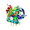 1z78S S: Starting model for refinement C: citing same article ( |
|---|---|
| Similar structure data |


-Links
 PDBj
PDBj


- Assembly
| Deposited unit | 
| ||||||||
|---|---|---|---|---|---|---|---|---|---|
| 1 | 
| ||||||||
| 2 | 
| ||||||||
| Unit cell |
| ||||||||
| Details | TSPN-1 is a monomer by itself. In presence of DP10, it forms a heparin-linked dimer. |
-Components
| #1: Protein | Mass: 27556.102 Da / Num. of mol.: 2 / Fragment: N-terminal domain Source method: isolated from a genetically manipulated source Source: (gene. exp.) Homo sapiens (human) / Gene: THBS1, TSP, TSP1 / Plasmid: PMT/BIP/V5-HIS A / Production host:  #2: Chemical | ChemComp-SO4 /   Mass: 96.063 Da / Num. of mol.: 6 / Source method: obtained synthetically / Formula: SO4 Mass: 96.063 Da / Num. of mol.: 6 / Source method: obtained synthetically / Formula: SO4#3: Water | ChemComp-HOH / |  Mass: 18.015 Da / Num. of mol.: 87 / Source method: isolated from a natural source / Formula: H2O Mass: 18.015 Da / Num. of mol.: 87 / Source method: isolated from a natural source / Formula: H2OHas protein modification | Y | |
|---|
-Experimental details
-Experiment
| Experiment | Method: X-RAY DIFFRACTION / Number of used crystals: 1 |
|---|
- Sample preparation
| Crystal | Density Matthews: 1.82 Å3/Da / Density % sol: 32.24 % |
|---|---|
| Crystal grow | Temperature: 293 K / Method: vapor diffusion, hanging drop / pH: 4.6 Details: 30% PEG1500, 0.08M Sodium acetate, pH 4.6, VAPOR DIFFUSION, HANGING DROP, temperature 293K |
-Data collection
| Diffraction | Mean temperature: 100 K |
|---|---|
| Diffraction source | Source: SYNCHROTRON / Site: APS  / Beamline: 19-ID / Wavelength: 0.99187 Å / Beamline: 19-ID / Wavelength: 0.99187 Å |
| Detector | Type: ADSC QUANTUM 315 / Detector: CCD / Date: Nov 2, 2004 / Details: Mirror |
| Radiation | Monochromator: Si 111 Crystal / Protocol: SINGLE WAVELENGTH / Monochromatic (M) / Laue (L): M / Scattering type: x-ray |
| Radiation wavelength | Wavelength: 0.99187 Å / Relative weight: 1 |
| Reflection | Resolution: 2.4→100 Å / Num. all: 14771 / Num. obs: 14771 / % possible obs: 88.8 % / Observed criterion σ(F): 0 / Observed criterion σ(I): 0 / Redundancy: 4.4 % / Rmerge(I) obs: 0.046 / Net I/σ(I): 29.87 |
| Reflection shell | Resolution: 2.4→2.49 Å / Redundancy: 2.8 % / Rmerge(I) obs: 0.268 / Mean I/σ(I) obs: 3.63 / Num. unique all: 1048 / % possible all: 65.9 |
- Processing
| Software |
| ||||||||||||||||||||
|---|---|---|---|---|---|---|---|---|---|---|---|---|---|---|---|---|---|---|---|---|---|
| Refinement | Method to determine structure: MOLECULAR REPLACEMENT Starting model: PDB entry 1Z78 Resolution: 2.4→100 Å / σ(F): 0 / σ(I): 0 Details: Fractionated heparin DP10 is chemically heterogeneous and partially disordered in the structure. Authors were able to identify only some SO4 groups from the heparin but not the heparin ...Details: Fractionated heparin DP10 is chemically heterogeneous and partially disordered in the structure. Authors were able to identify only some SO4 groups from the heparin but not the heparin backbone due to its possible mobility. Instead, there are several water molecules being positioned into some uncharacterized densities near heparin binding sites. These densities are likely from partially disordered heparin dp10, not necessarily from water molecules. Including of these waters in the model was just for the refinement purposes.
| ||||||||||||||||||||
| Refinement step | Cycle: LAST / Resolution: 2.4→100 Å
| ||||||||||||||||||||
| Refine LS restraints |
| ||||||||||||||||||||
| LS refinement shell | Resolution: 2.4→2.48 Å /
|
