 Movie
Movie Controller

[English] 日本語
 Yorodumi
- PDB-2os1: Structures of actinonin bound peptide deformylases from E. faecal... -
+ Open data

- Basic information
| Entry | Database: PDB / ID: 2os1 | ||||||
|---|---|---|---|---|---|---|---|
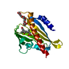
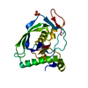
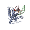
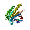
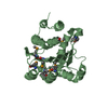
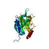
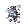
| Title | Structures of actinonin bound peptide deformylases from E. faecalis and S. pyogenes | ||||||
 Components Components | Peptide deformylase | ||||||
 Keywords Keywords | HYDROLASE / PDF / peptide deformylase | ||||||
| Function / homology |  Function and homology information Function and homology informationpeptide deformylase / peptide deformylase activity / translation / metal ion binding Similarity search - Function | ||||||
| Biological species |   Enterococcus faecalis (bacteria) Enterococcus faecalis (bacteria) | ||||||
| Method |  X-RAY DIFFRACTION / SYNCHROTRON / MOLECULAR REPLACEMENT / Resolution: 1.5 Å X-RAY DIFFRACTION / SYNCHROTRON / MOLECULAR REPLACEMENT / Resolution: 1.5 Å | ||||||
 Authors Authors | Kim, E.E. / Kim, K.-H. / Moon, J.H. / Choi, K. / Lee, H.K. / Park, H.S. | ||||||
 Citation Citation | Journal: To be Published Title: Structures of actinonin bound peptide deformylases from E. faecalis and S. pyogenes Authors: Kim, E.E. / Kim, K.-H. / Moon, J.H. / Choi, K. / Lee, H.K. / Park, H.S. | ||||||
| History |
|
- Structure visualization
| Structure viewer | Molecule: MolmilJmol/JSmol |
|---|
- Downloads & links
-Download
| PDBx/mmCIF format | 2os1.cif.gz | 55.8 KB | Display | PDBx/mmCIF format |
|---|---|---|---|---|
| PDB format | pdb2os1.ent.gz | 38.8 KB | Display | PDB format |
| PDBx/mmJSON format | 2os1.json.gz | Tree view | PDBx/mmJSON format | |
| Others |  Other downloads Other downloads |
-Validation report
| Arichive directory | https://data.pdbj.org/pub/pdb/validation_reports/os/2os1ftp://data.pdbj.org/pub/pdb/validation_reports/os/2os1 | HTTPS FTP |
|---|
-Related structure data
| Related structure data |  2os0C 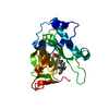 2os3C  1lqyS C: citing same article ( S: Starting model for refinement |
|---|---|
| Similar structure data |


-Links
 PDBj
PDBj- Assembly
| Deposited unit | 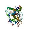
| ||||||||
|---|---|---|---|---|---|---|---|---|---|
| 1 |
| ||||||||
| Unit cell |
|
-Components
| #1: Protein | Mass: 21051.033 Da / Num. of mol.: 1 Source method: isolated from a genetically manipulated source Source: (gene. exp.) Enterococcus faecalis (bacteria) / Strain: ATCC 700802 / Gene: def / Plasmid: pET22b / Species (production host): Escherichia coli / Production host: | ||||
|---|---|---|---|---|---|
| #2: Chemical | ChemComp-NI /   Mass: 58.693 Da / Num. of mol.: 1 / Source method: obtained synthetically / Formula: Ni Mass: 58.693 Da / Num. of mol.: 1 / Source method: obtained synthetically / Formula: Ni | ||||
| #3: Chemical |   Mass: 96.063 Da / Num. of mol.: 3 / Source method: obtained synthetically / Formula: SO4 Mass: 96.063 Da / Num. of mol.: 3 / Source method: obtained synthetically / Formula: SO4#4: Chemical | ChemComp-BB2 / |   Mass: 385.498 Da / Num. of mol.: 1 / Source method: obtained synthetically / Formula: C19H35N3O5 / Comment: antitumor, antibiotic*YM Mass: 385.498 Da / Num. of mol.: 1 / Source method: obtained synthetically / Formula: C19H35N3O5 / Comment: antitumor, antibiotic*YM#5: Water | ChemComp-HOH / |  Mass: 18.015 Da / Num. of mol.: 210 / Source method: isolated from a natural source / Formula: H2O Mass: 18.015 Da / Num. of mol.: 210 / Source method: isolated from a natural source / Formula: H2O |
-Experimental details
-Experiment
| Experiment | Method: X-RAY DIFFRACTION / Number of used crystals: 1 |
|---|
- Sample preparation
| Crystal | Density Matthews: 2.26 Å3/Da / Density % sol: 45.51 % |
|---|---|
| Crystal grow | Temperature: 294 K / Method: vapor diffusion, hanging drop / pH: 7.4 Details: 16% PEG 8000, 0.2M Ammonium sulfate, pH 7.4, VAPOR DIFFUSION, HANGING DROP, temperature 294K |
-Data collection
| Diffraction | Mean temperature: 100 K |
|---|---|
| Diffraction source | Source: SYNCHROTRON / Site: PAL/PLS  / Beamline: 6B / Wavelength: 1.12714 Å / Beamline: 6B / Wavelength: 1.12714 Å |
| Detector | Type: BRUKER PROTEUM 300 / Detector: CCD / Date: Nov 26, 2004 / Details: mirrors |
| Radiation | Monochromator: DOUBLE CRYSTAL / Protocol: SINGLE WAVELENGTH / Monochromatic (M) / Laue (L): M / Scattering type: x-ray |
| Radiation wavelength | Wavelength: 1.12714 Å / Relative weight: 1 |
| Reflection | Resolution: 1.4→50 Å / Num. obs: 37104 / % possible obs: 86.4 % / Observed criterion σ(F): 0 / Observed criterion σ(I): 0 / Biso Wilson estimate: 11.9 Å2 / Rmerge(I) obs: 0.052 / Net I/σ(I): 28.3 |
| Reflection shell | Resolution: 1.4→1.45 Å / Rmerge(I) obs: 0.283 / Mean I/σ(I) obs: 4.5 / % possible all: 19.6 |
- Processing
| Software |
| ||||||||||||||||||||||||||||||||||||
|---|---|---|---|---|---|---|---|---|---|---|---|---|---|---|---|---|---|---|---|---|---|---|---|---|---|---|---|---|---|---|---|---|---|---|---|---|---|
| Refinement | Method to determine structure: MOLECULAR REPLACEMENT Starting model: PDB ENTRY 1LQY Resolution: 1.5→24.04 Å / Rfactor Rfree error: 0.005 / Data cutoff high absF: 336726.92 / Data cutoff low absF: 0 / Isotropic thermal model: RESTRAINED / Cross valid method: THROUGHOUT / σ(F): 0
| ||||||||||||||||||||||||||||||||||||
| Solvent computation | Solvent model: FLAT MODEL / Bsol: 45.0861 Å2 / ksol: 0.377374 e/Å3 | ||||||||||||||||||||||||||||||||||||
| Displacement parameters | Biso mean: 13.2 Å2
| ||||||||||||||||||||||||||||||||||||
| Refine analyze |
| ||||||||||||||||||||||||||||||||||||
| Refinement step | Cycle: LAST / Resolution: 1.5→24.04 Å
| ||||||||||||||||||||||||||||||||||||
| Refine LS restraints |
| ||||||||||||||||||||||||||||||||||||
| LS refinement shell | Resolution: 1.5→1.59 Å / Rfactor Rfree error: 0.015 / Total num. of bins used: 6
| ||||||||||||||||||||||||||||||||||||
| Xplor file |
|
