 Movie
Movie Controller

[English] 日本語
 Yorodumi
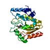
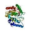
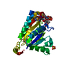
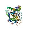
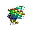
- PDB-3w9r: Crystal structure of the high-affinity abscisic acid receptor PYL... -
+ Open data

- Basic information
| Entry | Database: PDB / ID: 3w9r | ||||||
|---|---|---|---|---|---|---|---|
| Title | Crystal structure of the high-affinity abscisic acid receptor PYL9/RCAR9 bound to ABA | ||||||
 Components Components | Abscisic acid receptor PYL9 | ||||||
 Keywords Keywords | HORMONE RECEPTOR / Abscisic acid receptor / Drought tolerance / protein phosphatase inhibitor / START/Bet v1 family | ||||||
| Function / homology |  Function and homology information Function and homology informationabscisic acid binding / abscisic acid-activated signaling pathway / protein phosphatase inhibitor activity / signaling receptor activity / protein homodimerization activity / nucleus / plasma membrane / cytoplasm Similarity search - Function | ||||||
| Biological species |  | ||||||
| Method |  X-RAY DIFFRACTION / SYNCHROTRON / MOLECULAR REPLACEMENT / Resolution: 1.9 Å X-RAY DIFFRACTION / SYNCHROTRON / MOLECULAR REPLACEMENT / Resolution: 1.9 Å | ||||||
 Authors Authors | Nakagawa, M. / Hirano, Y. / Kagiyama, M. / Shibata, N. / Hakoshima, T. | ||||||
 Citation Citation | Journal: Genes Cells / Year: 2014 Title: Mechanism of high-affinity abscisic acid binding to PYL9/RCAR1. Authors: Nakagawa, M. / Kagiyama, M. / Shibata, N. / Hirano, Y. / Hakoshima, T. | ||||||
| History |
|
- Structure visualization
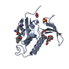
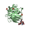
| Structure viewer | Molecule: MolmilJmol/JSmol |
|---|
- Downloads & links
-Download
| PDBx/mmCIF format | 3w9r.cif.gz | 84.8 KB | Display | PDBx/mmCIF format |
|---|---|---|---|---|
| PDB format | pdb3w9r.ent.gz | 63.2 KB | Display | PDB format |
| PDBx/mmJSON format | 3w9r.json.gz | Tree view | PDBx/mmJSON format | |
| Others |  Other downloads Other downloads |
-Validation report
| Arichive directory | https://data.pdbj.org/pub/pdb/validation_reports/w9/3w9rftp://data.pdbj.org/pub/pdb/validation_reports/w9/3w9r | HTTPS FTP |
|---|
-Related structure data
| Related structure data |  3cnwS S: Starting model for refinement |
|---|---|
| Similar structure data |


-Links
 PDBj
PDBj- Assembly
| Deposited unit | 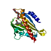
| ||||||||
|---|---|---|---|---|---|---|---|---|---|
| 1 |
| ||||||||
| Unit cell |
|
-Components
| #1: Protein | Mass: 21083.080 Da / Num. of mol.: 1 Source method: isolated from a genetically manipulated source Source: (gene. exp.)  |
|---|---|
| #2: Chemical | ChemComp-A8S / (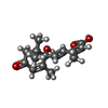  Mass: 264.317 Da / Num. of mol.: 1 / Source method: obtained synthetically / Formula: C15H20O4 / Comment: hormone*YM Mass: 264.317 Da / Num. of mol.: 1 / Source method: obtained synthetically / Formula: C15H20O4 / Comment: hormone*YM |
| #3: Chemical | ChemComp-P6G /   Mass: 282.331 Da / Num. of mol.: 1 / Source method: obtained synthetically / Formula: C12H26O7 / Comment: precipitant*YM Mass: 282.331 Da / Num. of mol.: 1 / Source method: obtained synthetically / Formula: C12H26O7 / Comment: precipitant*YM |
| #4: Water | ChemComp-HOH /  Mass: 18.015 Da / Num. of mol.: 70 / Source method: isolated from a natural source / Formula: H2O Mass: 18.015 Da / Num. of mol.: 70 / Source method: isolated from a natural source / Formula: H2O |
-Experimental details
-Experiment
| Experiment | Method: X-RAY DIFFRACTION / Number of used crystals: 1 |
|---|
- Sample preparation
| Crystal | Density Matthews: 3.44 Å3/Da / Density % sol: 64.24 % |
|---|---|
| Crystal grow | Temperature: 293 K / Method: vapor diffusion / pH: 7 Details: 16.5 (w/v)% PEG 1500, maric acid-MES-Tris (MMT) buffer, pH 7.0, VAPOR DIFFUSION, temperature 293K |
-Data collection
| Diffraction | Mean temperature: 100 K |
|---|---|
| Diffraction source | Source: SYNCHROTRON / Site: SPring-8  / Beamline: BL41XU / Wavelength: 1 Å / Beamline: BL41XU / Wavelength: 1 Å |
| Detector | Type: ADSC QUANTUM 315 / Detector: CCD / Date: Oct 19, 2009 / Details: mirrors |
| Radiation | Monochromator: Rotated-inclined double-crystal monochromator , Si (111) Protocol: SINGLE WAVELENGTH / Monochromatic (M) / Laue (L): M / Scattering type: x-ray |
| Radiation wavelength | Wavelength: 1 Å / Relative weight: 1 |
| Reflection | Resolution: 1.9→50 Å / Num. all: 23060 / Num. obs: 23009 / % possible obs: 99.8 % / Observed criterion σ(F): 0 / Observed criterion σ(I): 0 / Redundancy: 10.7 % / Rmerge(I) obs: 0.05 / Net I/σ(I): 60.7 |
| Reflection shell | Resolution: 1.9→1.97 Å / Redundancy: 10.4 % / Rmerge(I) obs: 0.57 / Mean I/σ(I) obs: 4.73 / Num. unique all: 2282 / % possible all: 100 |
- Processing
| Software |
| ||||||||||||||||||||||||||||||||||||||||||||||||||||||||||||||||||||||||||||||||||||||||||||||||||||||||||||||||||||||||||||||||||||||||||||||||||||||||||||||||||||||||||
|---|---|---|---|---|---|---|---|---|---|---|---|---|---|---|---|---|---|---|---|---|---|---|---|---|---|---|---|---|---|---|---|---|---|---|---|---|---|---|---|---|---|---|---|---|---|---|---|---|---|---|---|---|---|---|---|---|---|---|---|---|---|---|---|---|---|---|---|---|---|---|---|---|---|---|---|---|---|---|---|---|---|---|---|---|---|---|---|---|---|---|---|---|---|---|---|---|---|---|---|---|---|---|---|---|---|---|---|---|---|---|---|---|---|---|---|---|---|---|---|---|---|---|---|---|---|---|---|---|---|---|---|---|---|---|---|---|---|---|---|---|---|---|---|---|---|---|---|---|---|---|---|---|---|---|---|---|---|---|---|---|---|---|---|---|---|---|---|---|---|---|---|
| Refinement | Method to determine structure: MOLECULAR REPLACEMENT Starting model: 3CNW Resolution: 1.9→50 Å / Cor.coef. Fo:Fc: 0.958 / Cor.coef. Fo:Fc free: 0.953 / SU B: 4.156 / SU ML: 0.064 / Cross valid method: THROUGHOUT / σ(F): 0 / ESU R: 0.11 / ESU R Free: 0.102 / Stereochemistry target values: MAXIMUM LIKELIHOOD / Details: HYDROGENS HAVE BEEN USED IF PRESENT IN THE INPUT
| ||||||||||||||||||||||||||||||||||||||||||||||||||||||||||||||||||||||||||||||||||||||||||||||||||||||||||||||||||||||||||||||||||||||||||||||||||||||||||||||||||||||||||
| Solvent computation | Ion probe radii: 0.8 Å / Shrinkage radii: 0.8 Å / VDW probe radii: 1.2 Å / Solvent model: MASK | ||||||||||||||||||||||||||||||||||||||||||||||||||||||||||||||||||||||||||||||||||||||||||||||||||||||||||||||||||||||||||||||||||||||||||||||||||||||||||||||||||||||||||
| Displacement parameters | Biso mean: 40.305 Å2
| ||||||||||||||||||||||||||||||||||||||||||||||||||||||||||||||||||||||||||||||||||||||||||||||||||||||||||||||||||||||||||||||||||||||||||||||||||||||||||||||||||||||||||
| Refinement step | Cycle: LAST / Resolution: 1.9→50 Å
| ||||||||||||||||||||||||||||||||||||||||||||||||||||||||||||||||||||||||||||||||||||||||||||||||||||||||||||||||||||||||||||||||||||||||||||||||||||||||||||||||||||||||||
| Refine LS restraints |
| ||||||||||||||||||||||||||||||||||||||||||||||||||||||||||||||||||||||||||||||||||||||||||||||||||||||||||||||||||||||||||||||||||||||||||||||||||||||||||||||||||||||||||
| LS refinement shell | Resolution: 1.9→1.949 Å / Total num. of bins used: 20
| ||||||||||||||||||||||||||||||||||||||||||||||||||||||||||||||||||||||||||||||||||||||||||||||||||||||||||||||||||||||||||||||||||||||||||||||||||||||||||||||||||||||||||
| Refinement TLS params. | Method: refined / Origin x: 37.2245 Å / Origin y: -36.3593 Å / Origin z: -7.3056 Å
|
