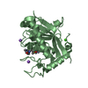
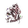
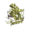
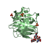
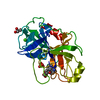
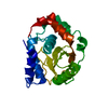
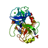
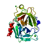
[English] 日本語
Yorodumi
- PDB-4dd8: ADAM-8 metalloproteinase domain with bound batimastat -

+
Open data


ID or keywords:

Loading...

-
Basic information

Entry
Database: PDB / ID: 4dd8
TitleADAM-8 metalloproteinase domain with bound batimastat
ComponentsDisintegrin and metalloproteinase domain-containing protein 8
KeywordsHYDROLASE/HYDROLASE INHIBITOR / batimastat / inflammation / alpha/beta motif / metalloproteinase / allergic asthma / tumorigenesis / arthritis / aberrant neural cell signaling / HYDROLASE-HYDROLASE INHIBITOR complex
Function / homology
Function and homology information


positive regulation of fibronectin-dependent thymocyte migration / positive regulation of eosinophil migration / dense core granule membrane / tertiary granule / positive regulation of tumor necrosis factor (ligand) superfamily member 11 production / positive regulation of neutrophil extravasation / phagolysosome / neutrophil degranulation / positive regulation of T cell differentiation in thymus / regulation of cell-cell adhesion ...positive regulation of fibronectin-dependent thymocyte migration / positive regulation of eosinophil migration / dense core granule membrane / tertiary granule / positive regulation of tumor necrosis factor (ligand) superfamily member 11 production / positive regulation of neutrophil extravasation / phagolysosome / neutrophil degranulation / positive regulation of T cell differentiation in thymus / regulation of cell-cell adhesion / lymphocyte chemotaxis / alpha9-beta1 integrin-ADAM8 complex / positive regulation of protein processing / leukocyte migration involved in inflammatory response / positive regulation of acute inflammatory response / specific granule / positive regulation of innate immune response / Hydrolases; Acting on peptide bonds (peptidases); Metalloendopeptidases / podosome / positive regulation of membrane protein ectodomain proteolysis / positive regulation of thymocyte apoptotic process / plasma membrane => GO:0005886 / tertiary granule membrane / ficolin-1-rich granule membrane / positive regulation of cell adhesion / extracellular matrix disassembly / positive regulation of bone resorption / specific granule membrane / cell adhesion molecule binding / Degradation of the extracellular matrix / : / positive regulation of protein secretion / positive regulation of MAP kinase activity / cell morphogenesis / metalloendopeptidase activity / cell-cell adhesion / metallopeptidase activity / positive regulation of NF-kappaB transcription factor activity / cellular response to hypoxia / angiogenesis / negative regulation of neuron apoptotic process / positive regulation of phosphatidylinositol 3-kinase/protein kinase B signal transduction / inflammatory response / serine-type endopeptidase activity / calcium ion binding / Neutrophil degranulation / cell surface / zinc ion binding / plasma membrane / cytoplasm
Similarity search - Function
ADAM cysteine-rich / ADAM, cysteine-rich domain / ADAM Cysteine-Rich Domain / Disintegrin, conserved site / Disintegrins signature. / Peptidase M12B, propeptide / Reprolysin family propeptide / Reprolysin domain, adamalysin-type / Reprolysin (M12B) family zinc metalloprotease / Disintegrin ...ADAM cysteine-rich / ADAM, cysteine-rich domain / ADAM Cysteine-Rich Domain / Disintegrin, conserved site / Disintegrins signature. / Peptidase M12B, propeptide / Reprolysin family propeptide / Reprolysin domain, adamalysin-type / Reprolysin (M12B) family zinc metalloprotease / Disintegrin / Disintegrin domain profile. / Homologues of snake disintegrins / Disintegrin domain / Disintegrin domain superfamily / Peptidase M12B, ADAM/reprolysin / ADAM type metalloprotease domain profile. / Collagenase (Catalytic Domain) / Collagenase (Catalytic Domain) / Metallopeptidase, catalytic domain superfamily / EGF-like domain profile. / EGF-like domain signature 2. / EGF-like domain / Neutral zinc metallopeptidases, zinc-binding region signature. / 3-Layer(aba) Sandwich / Alpha Beta
Similarity search - Domain/homology
Chem-BAT / : / Disintegrin and metalloproteinase domain-containing protein 8
Similarity search - Component
Biological speciesHomo sapiens (human)
MethodX-RAY DIFFRACTION / SYNCHROTRON / MOLECULAR REPLACEMENT / Resolution: 2.1 Å
AuthorsHall, T. / Shieh, H.S. / Day, J.E. / Caspers, N. / Chrencik, J.E. / Williams, J.M. / Pegg, L.E. / Pauley, A.M. / Moon, A.F. / Krahn, J.M. ...Hall, T. / Shieh, H.S. / Day, J.E. / Caspers, N. / Chrencik, J.E. / Williams, J.M. / Pegg, L.E. / Pauley, A.M. / Moon, A.F. / Krahn, J.M. / Fischer, D.H. / Kiefer, J.R. / Tomasselli, A.G. / Zack, M.D.
CitationJournal: Acta Crystallogr.,Sect.F / Year: 2012
Title: Structure of human ADAM-8 catalytic domain complexed with batimastat.
Authors: Hall, T. / Shieh, H.S. / Day, J.E. / Caspers, N. / Chrencik, J.E. / Williams, J.M. / Pegg, L.E. / Pauley, A.M. / Moon, A.F. / Krahn, J.M. / Fischer, D.H. / Kiefer, J.R. / Tomasselli, A.G. / Zack, M.D.
History
DepositionJan 18, 2012Deposition site: RCSB / Processing site: RCSB
Revision 1.0Jun 6, 2012Provider: repository / Type: Initial release
Revision 1.1Jun 27, 2012Group: Database references
Revision 1.2Sep 13, 2023Group: Data collection / Database references ...Data collection / Database references / Derived calculations / Refinement description
Category: chem_comp_atom / chem_comp_bond ...chem_comp_atom / chem_comp_bond / database_2 / pdbx_initial_refinement_model / pdbx_struct_conn_angle / struct_conn / struct_site
Item: _database_2.pdbx_DOI / _database_2.pdbx_database_accession ..._database_2.pdbx_DOI / _database_2.pdbx_database_accession / _pdbx_struct_conn_angle.ptnr1_auth_asym_id / _pdbx_struct_conn_angle.ptnr1_auth_comp_id / _pdbx_struct_conn_angle.ptnr1_auth_seq_id / _pdbx_struct_conn_angle.ptnr1_label_asym_id / _pdbx_struct_conn_angle.ptnr1_label_atom_id / _pdbx_struct_conn_angle.ptnr1_label_comp_id / _pdbx_struct_conn_angle.ptnr1_label_seq_id / _pdbx_struct_conn_angle.ptnr2_auth_asym_id / _pdbx_struct_conn_angle.ptnr2_auth_comp_id / _pdbx_struct_conn_angle.ptnr2_auth_seq_id / _pdbx_struct_conn_angle.ptnr2_label_asym_id / _pdbx_struct_conn_angle.ptnr2_label_atom_id / _pdbx_struct_conn_angle.ptnr2_label_comp_id / _pdbx_struct_conn_angle.ptnr3_auth_asym_id / _pdbx_struct_conn_angle.ptnr3_auth_comp_id / _pdbx_struct_conn_angle.ptnr3_auth_seq_id / _pdbx_struct_conn_angle.ptnr3_label_asym_id / _pdbx_struct_conn_angle.ptnr3_label_atom_id / _pdbx_struct_conn_angle.ptnr3_label_comp_id / _pdbx_struct_conn_angle.ptnr3_label_seq_id / _pdbx_struct_conn_angle.value / _struct_conn.pdbx_dist_value / _struct_conn.ptnr1_auth_asym_id / _struct_conn.ptnr1_auth_comp_id / _struct_conn.ptnr1_auth_seq_id / _struct_conn.ptnr1_label_asym_id / _struct_conn.ptnr1_label_atom_id / _struct_conn.ptnr1_label_comp_id / _struct_conn.ptnr1_label_seq_id / _struct_conn.ptnr2_auth_asym_id / _struct_conn.ptnr2_auth_comp_id / _struct_conn.ptnr2_auth_seq_id / _struct_conn.ptnr2_label_asym_id / _struct_conn.ptnr2_label_atom_id / _struct_conn.ptnr2_label_comp_id / _struct_site.pdbx_auth_asym_id / _struct_site.pdbx_auth_comp_id / _struct_site.pdbx_auth_seq_id

-
Structure visualization

Structure viewerMolecule:
MolmilJmol/JSmol

Downloads & links

-
Assembly

Deposited unit
A: Disintegrin and metalloproteinase domain-containing protein 8
B: Disintegrin and metalloproteinase domain-containing protein 8
C: Disintegrin and metalloproteinase domain-containing protein 8
D: Disintegrin and metalloproteinase domain-containing protein 8
hetero molecules


Theoretical massNumber of molelcules
Total (without water)96,15430
Polymers93,4174
Non-polymers2,73626
Water7,476415
1
A: Disintegrin and metalloproteinase domain-containing protein 8
hetero molecules


Theoretical massNumber of molelcules
Total (without water)24,0979
Polymers23,3541
Non-polymers7438
Water181
TypeNameSymmetry operationNumber
identity operation1_555x,y,z1
MethodPISA
2
B: Disintegrin and metalloproteinase domain-containing protein 8
hetero molecules


Theoretical massNumber of molelcules
Total (without water)24,0197
Polymers23,3541
Non-polymers6656
Water181
TypeNameSymmetry operationNumber
identity operation1_555x,y,z1
MethodPISA
3
C: Disintegrin and metalloproteinase domain-containing protein 8
hetero molecules


Theoretical massNumber of molelcules
Total (without water)24,0197
Polymers23,3541
Non-polymers6656
Water181
TypeNameSymmetry operationNumber
identity operation1_555x,y,z1
MethodPISA
4
D: Disintegrin and metalloproteinase domain-containing protein 8
hetero molecules


Theoretical massNumber of molelcules
Total (without water)24,0197
Polymers23,3541
Non-polymers6656
Water181
TypeNameSymmetry operationNumber
identity operation1_555x,y,z1
MethodPISA
Unit cell
Length a, b, c (Å)91.600, 50.900, 93.500
Angle α, β, γ (deg.)90.00, 102.40, 90.00
Int Tables number4
Space group name H-MP1211
Noncrystallographic symmetry (NCS)NCS domain:
IDEns-ID
11
21
31
41
12
22
32
42
13
23
33
43
14
24
34
44
15
25

NCS domain segments:
Dom-IDComponent-IDEns-IDSelection details
111chain A and (((resid 197:219 or resid 221:252 or resid...
211chain B and (((resid 197:219 or resid 221:252 or resid...
311chain C and (((resid 197:219 or resid 221:252 or resid...
411chain D and (((resid 197:219 or resid 221:252 or resid...
112chain A and resid 253:257 and backbone
212chain B and resid 253:257 and backbone
312chain C and resid 253:257 and backbone
412chain D and resid 253:257 and backbone
113chain A and resid 258:262
213chain B and resid 258:262
313chain C and resid 258:262
413chain D and resid 258:262
114chain A and resid 263:278 and backbone
214chain B and resid 263:278 and backbone
314chain C and resid 263:278 and backbone
414chain D and resid 263:278 and backbone
115chain B and resid 276:283
215chain D and resid 276:283

NCS ensembles :
ID
1
2
3
4
5

-
Components

-
Protein , 1 types, 4 molecules ABCD

#1: Protein
Disintegrin and metalloproteinase domain-containing protein 8 / ADAM 8 / Cell surface antigen MS2


Mass: 23354.297 Da / Num. of mol.: 4 / Fragment: UNP Residues 196-403
Source method: isolated from a genetically manipulated source
Source: (gene. exp.) Homo sapiens (human) / Gene: ADAM8, MS2 / Plasmid: pcDNAmychisB / Cell line (production host): HEK-293 Freestyle / Production host: Homo sapiens (human)
References: UniProt: P78325, Hydrolases; Acting on peptide bonds (peptidases); Metalloendopeptidases

-
Non-polymers , 7 types, 441 molecules

#2: Chemical
ChemComp-BAT / 4-(N-HYDROXYAMINO)-2R-ISOBUTYL-2S-(2-THIENYLTHIOMETHYL)SUCCINYL-L-PHENYLALANINE-N-METHYLAMIDE / BATIMASTAT / BB94


Mass: 477.640 Da / Num. of mol.: 4 / Source method: obtained synthetically / Formula: C23H31N3O4S2 / Comment: inhibitor*YM
#3: Chemical
ChemComp-CA / CALCIUM ION


Mass: 40.078 Da / Num. of mol.: 4 / Source method: obtained synthetically / Formula: Ca
#4: Chemical
ChemComp-ZN / ZINC ION


Mass: 65.409 Da / Num. of mol.: 4 / Source method: obtained synthetically / Formula: Zn
#5: Chemical
ChemComp-NA / SODIUM ION


Mass: 22.990 Da / Num. of mol.: 8 / Source method: obtained synthetically / Formula: Na
#6: Chemical
ChemComp-CL / CHLORIDE ION


Mass: 35.453 Da / Num. of mol.: 4 / Source method: obtained synthetically / Formula: Cl
#7: Chemical ChemComp-K / POTASSIUM ION


Mass: 39.098 Da / Num. of mol.: 2 / Source method: obtained synthetically / Formula: K
#8: Water ChemComp-HOH / water


Mass: 18.015 Da / Num. of mol.: 415 / Source method: isolated from a natural source / Formula: H2O

-
Experimental details

-
Experiment

ExperimentMethod: X-RAY DIFFRACTION / Number of used crystals: 1

-
Sample preparation

CrystalDensity Matthews: 2.28 Å3/Da / Density % sol: 46.03 %
Crystal growTemperature: 291 K / Method: vapor diffusion, sitting drop / pH: 8
Details: Purified ADAM-8/batimastat complex (10mg/mL) was crystallized by the sitting-drop vapor diffusion method, mixing 1uL complex with 1uL of 0.1M Tris pH 8.0, 1M sodium formate, 10% methanol. ...Details: Purified ADAM-8/batimastat complex (10mg/mL) was crystallized by the sitting-drop vapor diffusion method, mixing 1uL complex with 1uL of 0.1M Tris pH 8.0, 1M sodium formate, 10% methanol. Crystals reached usable size in 10 days. Crystals were then transferred to a cryo solution containing 0.1M Tris pH 8.0, 1.2M sodium formate, 10% methanol, and 10% glycerol. The crystals were harvested and flash frozen in liquid nitrogen for data collection., VAPOR DIFFUSION, SITTING DROP, temperature 291K

-
Data collection

DiffractionMean temperature: 100 K
Diffraction sourceSource: SYNCHROTRON / Site: ALS / Beamline: 5.0.1 / Wavelength: 1 Å
DetectorType: ADSC QUANTUM 315r / Detector: CCD / Details: mirrors
RadiationMonochromator: Si(220) Asymmetric cut single crystal / Protocol: SINGLE WAVELENGTH / Monochromatic (M) / Laue (L): M / Scattering type: x-ray
Radiation wavelengthWavelength: 1 Å / Relative weight: 1
Reflection twinOperator: l,-k,h / Fraction: 0.26
ReflectionResolution: 2.1→50 Å / Num. all: 49827 / Num. obs: 49479 / % possible obs: 99.3 % / Observed criterion σ(F): -3 / Observed criterion σ(I): -3 / Redundancy: 3.6 % / Rsym value: 0.106 / Net I/σ(I): 10.9
Reflection shellResolution: 2.1→2.18 Å / Redundancy: 2.8 % / Mean I/σ(I) obs: 2.92 / Num. unique all: 4746 / Rsym value: 0.365 / % possible all: 95.9

-
Processing

Software
NameVersionClassification
ADSCQuantumdata collection
MOLREPphasing
PHENIX(phenix.refine: 1.7_650)refinement
REFMACrefinement
HKL-2000data reduction
HKL-2000data scaling
RefinementMethod to determine structure: MOLECULAR REPLACEMENT
Starting model: PDB ENTRY 1R55
Resolution: 2.1→44.745 Å / Cross valid method: R-free / σ(F): 1 / Phase error: 36.91 / Stereochemistry target values: TWIN_LSQ_F
RfactorNum. reflection% reflectionSelection details
Rfree0.255 2368 4.97 %random
Rwork0.1881 ---
obs0.1916 47689 96.04 %-
all-49479 --
Solvent computationShrinkage radii: 0.83 Å / VDW probe radii: 1.1 Å / Solvent model: FLAT BULK SOLVENT MODEL / Bsol: 50.471 Å2 / ksol: 0.37 e/Å3
Displacement parameters
Baniso -1Baniso -2Baniso -3
1-3.3066 Å2-0 Å2-12.629 Å2
2---4.8035 Å20 Å2
3---1.4969 Å2
Refinement stepCycle: LAST / Resolution: 2.1→44.745 Å
ProteinNucleic acidLigandSolventTotal
Num. atoms6270 0 150 415 6835
Refine LS restraints
Refine-IDTypeDev idealNumber
X-RAY DIFFRACTIONf_bond_d0.0086718
X-RAY DIFFRACTIONf_angle_d0.6828982
X-RAY DIFFRACTIONf_dihedral_angle_d12.892340
X-RAY DIFFRACTIONf_chiral_restr0.049990
X-RAY DIFFRACTIONf_plane_restr0.0021200
Refine LS restraints NCS
Ens-IDDom-IDAuth asym-IDNumberRefine-IDTypeRms dev position (Å)
11A674X-RAY DIFFRACTIONPOSITIONAL
12B674X-RAY DIFFRACTIONPOSITIONAL0.155
13C682X-RAY DIFFRACTIONPOSITIONAL0.122
14D670X-RAY DIFFRACTIONPOSITIONAL0.151
21A20X-RAY DIFFRACTIONPOSITIONAL
22B20X-RAY DIFFRACTIONPOSITIONAL0.237
23C20X-RAY DIFFRACTIONPOSITIONAL0.127
24D20X-RAY DIFFRACTIONPOSITIONAL0.145
31A41X-RAY DIFFRACTIONPOSITIONAL
32B41X-RAY DIFFRACTIONPOSITIONAL0.138
33C41X-RAY DIFFRACTIONPOSITIONAL0.118
34D41X-RAY DIFFRACTIONPOSITIONAL0.145
41A60X-RAY DIFFRACTIONPOSITIONAL
42B60X-RAY DIFFRACTIONPOSITIONAL0.211
43C60X-RAY DIFFRACTIONPOSITIONAL0.187
44D60X-RAY DIFFRACTIONPOSITIONAL0.209
51B65X-RAY DIFFRACTIONPOSITIONAL
52D65X-RAY DIFFRACTIONPOSITIONAL0.078
LS refinement shell
Resolution (Å)Rfactor RfreeNum. reflection RfreeRfactor RworkNum. reflection RworkRefine-ID% reflection obs (%)
2.1-2.14290.3006970.23612354X-RAY DIFFRACTION82
2.1429-2.18940.28971180.22652506X-RAY DIFFRACTION87
2.1894-2.24030.26341200.22532514X-RAY DIFFRACTION87
2.2403-2.29630.32881220.22762554X-RAY DIFFRACTION88
2.2963-2.35830.27031460.2272589X-RAY DIFFRACTION90
2.3583-2.42760.29531410.21812653X-RAY DIFFRACTION91
2.4276-2.50590.29911460.20942619X-RAY DIFFRACTION91
2.5059-2.59530.30291330.21112709X-RAY DIFFRACTION92
2.5953-2.69910.27081600.21242683X-RAY DIFFRACTION93
2.6991-2.82170.28061500.20382700X-RAY DIFFRACTION93
2.8217-2.97020.3241260.22745X-RAY DIFFRACTION94
2.9702-3.15580.2571150.1842764X-RAY DIFFRACTION95
3.1558-3.39880.25681270.17142783X-RAY DIFFRACTION95
3.3988-3.73950.22431430.16462749X-RAY DIFFRACTION94
3.7395-4.27750.21961550.14662746X-RAY DIFFRACTION93
4.2775-5.37750.19231390.15142791X-RAY DIFFRACTION95
5.3775-22.83110.2491690.19452873X-RAY DIFFRACTION94

+
About Yorodumi

-
News

-
Feb 9, 2022. New format data for meta-information of EMDB entries

New format data for meta-information of EMDB entries

  • Version 3 of the EMDB header file is now the official format.
  • The previous official version 1.9 will be removed from the archive.

Related info.:EMDB header

External links:wwPDB to switch to version 3 of the EMDB data model

-
Aug 12, 2020. Covid-19 info

Covid-19 info

URL: https://pdbjlvh1.pdbj.org/emnavi/covid19.php

New page: Covid-19 featured information page in EM Navigator.

Related info.:Covid-19 info / Mar 5, 2020. Novel coronavirus structure data

+
Mar 5, 2020. Novel coronavirus structure data

Novel coronavirus structure data

Related info.:Yorodumi Speices / Aug 12, 2020. Covid-19 info

External links:COVID-19 featured content - PDBj / Molecule of the Month (242):Coronavirus Proteases

+
Jan 31, 2019. EMDB accession codes are about to change! (news from PDBe EMDB page)

EMDB accession codes are about to change! (news from PDBe EMDB page)

  • The allocation of 4 digits for EMDB accession codes will soon come to an end. Whilst these codes will remain in use, new EMDB accession codes will include an additional digit and will expand incrementally as the available range of codes is exhausted. The current 4-digit format prefixed with “EMD-” (i.e. EMD-XXXX) will advance to a 5-digit format (i.e. EMD-XXXXX), and so on. It is currently estimated that the 4-digit codes will be depleted around Spring 2019, at which point the 5-digit format will come into force.
  • The EM Navigator/Yorodumi systems omit the EMD- prefix.

Related info.:Q: What is EMD? / ID/Accession-code notation in Yorodumi/EM Navigator

External links:EMDB Accession Codes are Changing Soon! / Contact to PDBj

+
Jul 12, 2017. Major update of PDB

Major update of PDB

  • wwPDB released updated PDB data conforming to the new PDBx/mmCIF dictionary.
  • This is a major update changing the version number from 4 to 5, and with Remediation, in which all the entries are updated.
  • In this update, many items about electron microscopy experimental information are reorganized (e.g. em_software).
  • Now, EM Navigator and Yorodumi are based on the updated data.

External links:wwPDB Remediation / Enriched Model Files Conforming to OneDep Data Standards Now Available in the PDB FTP Archive

-
Yorodumi

Thousand views of thousand structures

  • Yorodumi is a browser for structure data from EMDB, PDB, SASBDB, etc.
  • This page is also the successor to EM Navigator detail page, and also detail information page/front-end page for Omokage search.
  • The word "yorodu" (or yorozu) is an old Japanese word meaning "ten thousand". "mi" (miru) is to see.

Related info.:EMDB / PDB / SASBDB / Comparison of 3 databanks / Yorodumi Search / Aug 31, 2016. New EM Navigator & Yorodumi / Yorodumi Papers / Jmol/JSmol / Function and homology information / Changes in new EM Navigator and Yorodumi

Read more