 Movie
Movie Controller

[English] 日本語
 Yorodumi
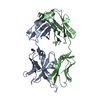
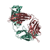
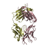
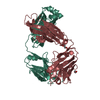
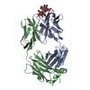
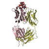
- PDB-2omn: Bence Jones KWR Protein- Immunoglobulin Light Chain Dimer, P4(3)2... -
+ Open data

- Basic information
| Entry | Database: PDB / ID: 2omn | |||||||||
|---|---|---|---|---|---|---|---|---|---|---|
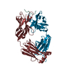
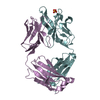
| Title | Bence Jones KWR Protein- Immunoglobulin Light Chain Dimer, P4(3)2(1)2 Crystal Form | |||||||||
 Components Components | Bence Jones KWR Protein - Immunoglobulin Light Chain | |||||||||
 Keywords Keywords | IMMUNE SYSTEM / Immunoglobulin Light Chain | |||||||||
| Function / homology | Immunoglobulins / Immunoglobulin-like / Sandwich / Mainly Beta / PHENOL Function and homology information Function and homology information | |||||||||
| Biological species |  Homo sapiens (human) Homo sapiens (human) | |||||||||
| Method |  X-RAY DIFFRACTION / SYNCHROTRON / MOLECULAR REPLACEMENT / Resolution: 2.2 Å X-RAY DIFFRACTION / SYNCHROTRON / MOLECULAR REPLACEMENT / Resolution: 2.2 Å | |||||||||
 Authors Authors | Makino, D.L. / Larson, S.B. / McPherson, A. | |||||||||
 Citation Citation | Journal: Acta Crystallogr.,Sect.D / Year: 2007 Title: Bence Jones KWR protein structures determined by X-ray crystallography. Authors: Makino, D.L. / Henschen-Edman, A.H. / Larson, S.B. / McPherson, A. | |||||||||
| History |
|
- Structure visualization
| Structure viewer | Molecule: MolmilJmol/JSmol |
|---|
- Downloads & links
-Download
| PDBx/mmCIF format | 2omn.cif.gz | 92.8 KB | Display | PDBx/mmCIF format |
|---|---|---|---|---|
| PDB format | pdb2omn.ent.gz | 71.9 KB | Display | PDB format |
| PDBx/mmJSON format | 2omn.json.gz | Tree view | PDBx/mmJSON format | |
| Others |  Other downloads Other downloads |
-Validation report
| Arichive directory | https://data.pdbj.org/pub/pdb/validation_reports/om/2omnftp://data.pdbj.org/pub/pdb/validation_reports/om/2omn | HTTPS FTP |
|---|
-Related structure data
| Related structure data | 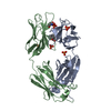 2oldC 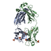 2ombC  1dclS C: citing same article ( S: Starting model for refinement |
|---|---|
| Similar structure data |


-Links
 PDBj
PDBj


- Assembly
| Deposited unit | 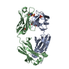
| ||||||||
|---|---|---|---|---|---|---|---|---|---|
| 1 |
| ||||||||
| Unit cell |
|
-Components
| #1: Antibody | Mass: 22698.914 Da / Num. of mol.: 2 / Source method: isolated from a natural source / Details: urine / Source: (natural) Homo sapiens (human) / Strain: KWR#2: Chemical |   Mass: 96.063 Da / Num. of mol.: 3 / Source method: obtained synthetically / Formula: SO4 Mass: 96.063 Da / Num. of mol.: 3 / Source method: obtained synthetically / Formula: SO4#3: Chemical | ChemComp-IPH / | 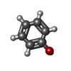  Mass: 94.111 Da / Num. of mol.: 1 / Source method: obtained synthetically / Formula: C6H6O Mass: 94.111 Da / Num. of mol.: 1 / Source method: obtained synthetically / Formula: C6H6O#4: Water | ChemComp-HOH / |  Mass: 18.015 Da / Num. of mol.: 98 / Source method: isolated from a natural source / Formula: H2O Mass: 18.015 Da / Num. of mol.: 98 / Source method: isolated from a natural source / Formula: H2OHas protein modification | Y | |
|---|
-Experimental details
-Experiment
| Experiment | Method: X-RAY DIFFRACTION / Number of used crystals: 1 |
|---|
- Sample preparation
| Crystal | Density Matthews: 2.35 Å3/Da / Density % sol: 47.75 % |
|---|---|
| Crystal grow | Temperature: 295 K / Method: vapor diffusion, sitting drop / pH: 8 Details: 1.5M Ammonium Sulfate, 0.1M Tris/HCl pH 8.0, VAPOR DIFFUSION, SITTING DROP, temperature 295K |
-Data collection
| Diffraction | Mean temperature: 100 K |
|---|---|
| Diffraction source | Source: SYNCHROTRON / Site: ALS  / Beamline: 5.0.2 / Wavelength: 1 Å / Beamline: 5.0.2 / Wavelength: 1 Å |
| Detector | Type: ADSC QUANTUM 210 / Detector: CCD / Date: Jul 11, 2003 |
| Radiation | Monochromator: Si(111) / Protocol: SINGLE WAVELENGTH / Monochromatic (M) / Laue (L): M / Scattering type: x-ray |
| Radiation wavelength | Wavelength: 1 Å / Relative weight: 1 |
| Reflection | Resolution: 2.08→46.88 Å / Num. all: 20595 / Num. obs: 20595 / % possible obs: 76.8 % / Observed criterion σ(F): 0 / Observed criterion σ(I): 0 / Redundancy: 9.8 % / Biso Wilson estimate: 35.1 Å2 / Limit h max: 32 / Limit h min: 0 / Limit k max: 18 / Limit k min: 0 / Limit l max: 73 / Limit l min: 0 / Observed criterion F max: 229355.06 / Observed criterion F min: 0.27 / Rsym value: 0.059 / Net I/σ(I): 23.1 |
| Reflection shell | Resolution: 2.08→2.16 Å / Redundancy: 1.7 % / Mean I/σ(I) obs: 2.5 / Num. unique all: 394 / Rsym value: 0.336 / % possible all: 15.1 |
- Processing
| Software |
| |||||||||||||||||||||||||||
|---|---|---|---|---|---|---|---|---|---|---|---|---|---|---|---|---|---|---|---|---|---|---|---|---|---|---|---|---|
| Refinement | Method to determine structure: MOLECULAR REPLACEMENT Starting model: PDB ENTRY 1DCL Resolution: 2.2→45 Å / Rfactor Rfree error: 0.006 / Occupancy max: 1 / Occupancy min: 1 / Isotropic thermal model: Isotropic / Cross valid method: THROUGHOUT / σ(F): 0 / σ(I): 0 / Stereochemistry target values: Engh & Huber Details: PARAMETERS FOR PYROGLUTAMIC ACID AND PHENOL WERE OBTAINED FROM HETERO-COMPOUND INFORMATION CENTRE - UPPSALA.
| |||||||||||||||||||||||||||
| Solvent computation | Solvent model: CNS bulk solvent model used / Bsol: 37.1035 Å2 / ksol: 0.337824 e/Å3 | |||||||||||||||||||||||||||
| Displacement parameters | Biso max: 132.05 Å2 / Biso mean: 58.22 Å2 / Biso min: 32.89 Å2
| |||||||||||||||||||||||||||
| Refine analyze |
| |||||||||||||||||||||||||||
| Refinement step | Cycle: LAST / Resolution: 2.2→45 Å
| |||||||||||||||||||||||||||
| Refine LS restraints |
| |||||||||||||||||||||||||||
| LS refinement shell | Resolution: 2.2→2.28 Å / Rfactor Rfree error: 0.038
| |||||||||||||||||||||||||||
| Xplor file |
|
